Recognition: unknown
Generative Modeling of Complex-Valued Brain MRI Data
Pith reviewed 2026-05-10 09:18 UTC · model grok-4.3
The pith
A generative model for complex-valued brain MRI produces synthetic scans that let classifiers detect abnormalities more accurately than training on real data alone.
A machine-rendered reading of the paper's core claim, the machinery that carries it, and where it could break.
Core claim
The framework uses a conditional variational autoencoder to compress complex-valued MRI into latent codes while preserving phase coherence above 0.997, then applies a flow-matching model to generate new complex scans. Real-versus-synthetic classifiers give AUROC scores between 0.50 and 0.66, showing the outputs are nearly indistinguishable from real data. Classifiers trained entirely on the synthetic data achieve an AUROC of 0.880 for normal-versus-abnormal classification on the fastMRI dataset, surpassing the 0.842 baseline obtained from real data, and this performance advantage carries over to an independent external test set with biopsy-confirmed labels.
What carries the argument
Conditional variational autoencoder that preserves phase coherence in complex MRI, paired with a flow-matching generative model that samples new magnitude-plus-phase images.
If this is right
- Synthetic complex-valued MRI can augment scarce real datasets for training diagnostic models without loss of performance.
- Phase data in MRI contains features useful for distinguishing normal from abnormal tissue that magnitude images alone miss.
- The same generative approach can supply labeled training examples for rare conditions where collecting real scans is difficult.
- Generated samples maintain utility across different scanners and institutions, supporting multi-site model development.
Where Pith is reading between the lines
- If phase truly improves detection, reconstruction software could be redesigned to output and store complex data rather than magnitude only.
- The same joint magnitude-phase modeling could be tested on other modalities that acquire complex signals, such as certain CT or ultrasound techniques.
- A controlled ablation that removes phase from the synthetic data and measures the resulting drop in classifier AUROC would isolate how much of the gain comes from phase versus the generative process.
Load-bearing premise
The phase information encoded and regenerated by the model carries genuine pathology signals that improve classification without introducing spurious patterns that happen to work only on the chosen test sets.
What would settle it
Retraining the downstream normal-versus-abnormal classifier on a larger, multi-institution real dataset and finding that its AUROC exceeds the synthetic-data AUROC of 0.880 would show the reported advantage does not generalize.
Figures


read the original abstract
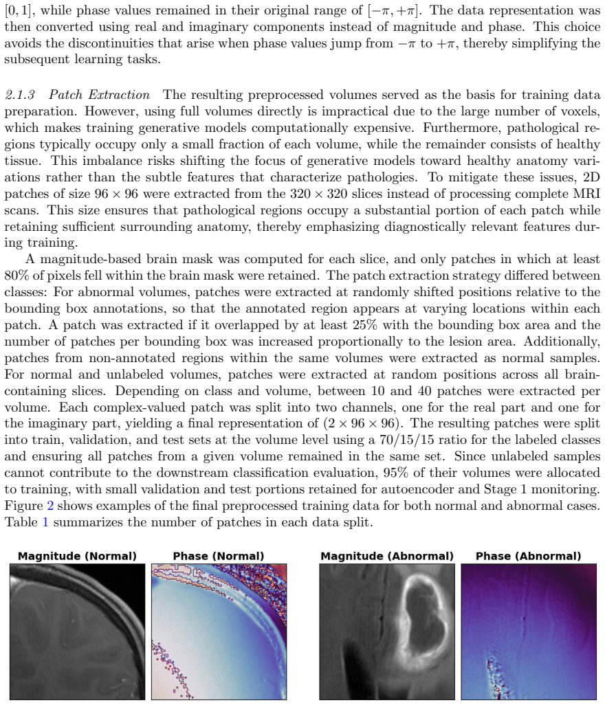
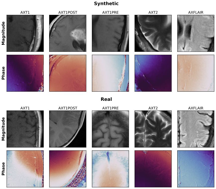
Objective. Standard Magnetic Resonance Imaging (MRI) reconstruction pipelines discard phase information captured during acquisition, despite evidence that it encodes tissue properties relevant to tumor diagnosis. Current machine learning approaches inherit this limitation by operating exclusively on reconstructed magnitude images. The aim of this study is to build a generative framework which is capable of jointly modeling magnitude and phase information of complex-valued MRI scans. Approach. The proposed generative framework combines a conditional variational autoencoder, which compresses complex-valued MRI scans into compact latent representations while preserving phase coherence, with a flow-matching-based generative model. Synthetic sample quality is assessed via a real-versus-synthetic classifier and by training downstream classifiers on synthetic data for abnormal tissue detection. Main results. The autoencoder preserves phase coherence above 0.997. Real-versus-synthetic classification yields low AUROC values between 0.50 and 0.66 across all acquisition sequences, indicating generated samples are nearly indistinguishable from real data. In downstream normal-versus-abnormal classification, classifiers trained entirely on synthetic data achieve an AUROC of 0.880, surpassing the real-data baseline of 0.842 on a publicly available dataset (fastMRI). This advantage persists on an independent external test set from a different institution with biopsy-confirmed labels. Significance. The proposed framework demonstrates the feasibility of jointly modeling magnitude and phase information for normal and abnormal complex-valued brain MRI data. Beyond synthetic data generation, it establishes a foundation for the usage of complete brain MRI information in future diagnostic applications and enables systematic investigation of how magnitude and phase jointly encode pathology-specific features.
Editorial analysis
A structured set of objections, weighed in public.
Referee Report
Summary. The manuscript proposes a generative framework that combines a conditional variational autoencoder (to compress complex-valued brain MRI while preserving phase) with a flow-matching model to synthesize realistic magnitude-and-phase data. It reports phase coherence above 0.997, real-versus-synthetic AUROCs of 0.50–0.66, and downstream normal-versus-abnormal classification AUROCs of 0.880 (synthetic) versus 0.842 (real) on fastMRI, with the advantage persisting on an external biopsy-labeled dataset from a different institution.
Significance. If the central empirical claims hold after verification, the work would be significant for demonstrating that joint modeling of magnitude and phase can produce synthetic data that improves downstream diagnostic performance beyond magnitude-only baselines. The use of a public dataset (fastMRI) and an independent external test set with biopsy confirmation strengthens the evaluation.
major comments (3)
- [Downstream evaluation] Downstream evaluation section: the AUROC gain (0.880 synthetic vs. 0.842 real) is presented as evidence that phase information contributes diagnostically relevant features, yet no ablation comparing complex-valued synthetic data against magnitude-only synthetic data is reported. This comparison is required to establish that the observed lift is attributable to phase modeling rather than other aspects of the generative pipeline.
- [External validation] External validation paragraph: the persistence of the performance advantage on the independent biopsy-labeled set is a key claim, but the manuscript provides no quantitative assessment of distribution shift (e.g., scanner parameters, acquisition protocols) between the generative training distribution and the external data. Without this, it remains possible that the reported edge reflects unmodeled covariates rather than genuine pathology encoding.
- [Methods] Methods, model architecture and training: the description of the conditional VAE latent dimension, conditioning variables, flow-matching network capacity, and training schedule is insufficient to reproduce the reported phase coherence >0.997 or to diagnose whether the downstream gains arise from faithful pathology encoding or from modeling artifacts.
minor comments (1)
- [Abstract] The abstract states real-versus-synthetic AUROC values as a range (0.50–0.66) without per-sequence breakdowns or reference to a supplementary table; adding this detail would improve clarity.
Simulated Author's Rebuttal
We thank the referee for the constructive comments, which highlight important aspects for strengthening our claims on phase-aware generative modeling of complex-valued MRI. We address each major comment below and will revise the manuscript to incorporate the requested analyses and details.
read point-by-point responses
-
Referee: [Downstream evaluation] Downstream evaluation section: the AUROC gain (0.880 synthetic vs. 0.842 real) is presented as evidence that phase information contributes diagnostically relevant features, yet no ablation comparing complex-valued synthetic data against magnitude-only synthetic data is reported. This comparison is required to establish that the observed lift is attributable to phase modeling rather than other aspects of the generative pipeline.
Authors: We agree that the current evidence would be strengthened by directly isolating the contribution of phase. In the revised manuscript we will add an ablation in which a magnitude-only generative model (trained and sampled without phase) is compared head-to-head with the complex-valued model on the same downstream abnormality-classification task. This will clarify whether the reported AUROC advantage arises from joint magnitude-phase modeling. revision: yes
-
Referee: [External validation] External validation paragraph: the persistence of the performance advantage on the independent biopsy-labeled set is a key claim, but the manuscript provides no quantitative assessment of distribution shift (e.g., scanner parameters, acquisition protocols) between the generative training distribution and the external data. Without this, it remains possible that the reported edge reflects unmodeled covariates rather than genuine pathology encoding.
Authors: We acknowledge the need for explicit quantification of distribution shift. In the revision we will add a dedicated paragraph reporting scanner field strengths, sequence parameters, and statistical measures of image-level shift (intensity histograms, contrast-to-noise ratios, and Kolmogorov-Smirnov tests) between the fastMRI training distribution and the external biopsy-labeled cohort. This analysis will help substantiate that the observed advantage is attributable to pathology encoding rather than acquisition covariates. revision: yes
-
Referee: [Methods] Methods, model architecture and training: the description of the conditional VAE latent dimension, conditioning variables, flow-matching network capacity, and training schedule is insufficient to reproduce the reported phase coherence >0.997 or to diagnose whether the downstream gains arise from faithful pathology encoding or from modeling artifacts.
Authors: We appreciate the call for greater reproducibility. The revised methods section will specify the cVAE latent dimension, the complete set of conditioning variables, the exact architecture and parameter count of the flow-matching network, and the full training schedule with all hyperparameters. These additions will enable independent reproduction of the phase-coherence results and allow readers to assess whether downstream gains stem from pathology encoding or modeling artifacts. revision: yes
Circularity Check
No circularity detected; results are empirical evaluations on held-out data
full rationale
The paper describes an empirical pipeline (conditional VAE + flow-matching) for generating complex-valued MRI and evaluates it via direct metrics: phase coherence >0.997, real-vs-synthetic AUROC 0.50-0.66, and downstream normal-vs-abnormal AUROC of 0.880 (synthetic-trained) vs 0.842 (real baseline) on fastMRI held-out data plus an external biopsy-labeled set. No mathematical derivation chain exists that reduces these quantities to fitted parameters by construction. No self-definitional steps, fitted-input predictions, or load-bearing self-citations are present in the reported results. The external test set and held-out real data provide independent benchmarks, keeping the evaluation self-contained without circular reduction.
Axiom & Free-Parameter Ledger
free parameters (2)
- latent dimension and conditioning variables
- flow-matching network capacity and training schedule
axioms (1)
- domain assumption The complex-valued MRI signal can be meaningfully compressed while preserving phase coherence for downstream pathology tasks.
Reference graph
Works this paper leans on
-
[1]
Siegel, Isabelle Soerjomataram, and Ahmedin Jemal
Freddie Bray, Mathieu Laversanne, Hyuna Sung, Jacques Ferlay, Rebecca L. Siegel, Isabelle Soerjomataram, and Ahmedin Jemal. Global cancer statistics 2022: GLOBOCAN estimates of incidence and mortality worldwide for 36 cancers in 185 countries.CA: A Cancer Journal for Clinicians, 74(3):229–263, 2024. https://doi.org/10.3322/caac.21834
-
[2]
Jakola, Thomas Skoglund, and Johan Ljungqvist
Anna Pennlund, Asgeir S. Jakola, Thomas Skoglund, and Johan Ljungqvist. A single-centre study of frame-based stereotactic brain biopsies.British Journal of Neurosurgery, 36(2): 213–216, 2022. https://doi.org/10.1080/02688697.2020.1867704
-
[3]
Luke Dixon, Gurpreet Kaur Jandu, Jai Sidpra, and Kshitij Mankad. Diagnostic accuracy of qualitative MRI in 550 paediatric brain tumours: evaluating current practice in the computational era.Quantitative Imaging in Medicine and Surgery, 12(1):131–143, 2022. https://doi.org/10.21037/qims-20-1388
-
[4]
Maximilien Riche, Aymeric Amelot, Matthieu Peyre, Laurent Capelle, Alexandre Carpentier, and Bertrand Mathon. Complications after frame-based stereotactic brain biopsy: a systematic review.Neurosurgical Review, 44(1):301–307, 2021. https://doi.org/10.1007/s10143-019-01234-w
-
[5]
Milica M. Badˇ za and MarkoˇC. Barjaktarovi´ c. Classification of brain tumors from MRI images using a convolutional neural network.Applied Sciences, 10(6):1999, 2020. https://doi.org/10.3390/app10061999. 14
-
[6]
Jihye Yun, Ji Eun Park, Hyunna Lee, Sungwon Ham, Namkug Kim, and Ho Sung Kim. Radiomic features and multilayer perceptron network classifier: A robust MRI classification strategy for distinguishing glioblastoma from primary central nervous system lymphoma. Scientific Reports, 9:5746, 2019. https://doi.org/10.1038/s41598-019-42276-w
-
[7]
Dunarea de Jos
Heba Mohsen, El-Sayed A. El-Dahshan, El-Sayed M. El-Horbaty, and Abdel-Badeeh M. Salem. Brain tumor type classification based on support vector machine in magnetic resonance images.Annals of the “Dunarea de Jos” University of Galati, Fascicle II, Mathematics, Physics, Theoretical Mechanics, 40(1), 2017
2017
-
[8]
Soheila Saeedi, Sorayya Rezayi, Hamidreza Keshavarz, and Sharareh R. Niakan Kalhori. MRI-based brain tumor detection using convolutional deep learning methods and chosen machine learning techniques.BMC Medical Informatics and Decision Making, 23(1):16, 2023. https://doi.org/10.1186/s12911-023-02114-6
-
[9]
Abdusalomov, Mukhriddin Mukhiddinov, and Taeg Keun Whangbo
Akmalbek B. Abdusalomov, Mukhriddin Mukhiddinov, and Taeg Keun Whangbo. Brain tumor detection based on deep learning approaches and magnetic resonance imaging.Cancers, 15(16):4172, 2023. https://doi.org/10.3390/cancers15164172
-
[10]
Mark Haacke, Yingbiao Xu, Yu-Chung N
E. Mark Haacke, Yingbiao Xu, Yu-Chung N. Cheng, and J¨ urgen R. Reichenbach. Susceptibility weighted imaging (SWI).Magnetic Resonance in Medicine, 52(3):612–618, 2004. https://doi.org/10.1002/mrm.20198
-
[11]
Ling-Wei Kong, Jin Chen, Heng Zhao, Kun Yao, Sheng-Yu Fang, Zheng Wang, Yin-Yan Wang, and Shou-Wei Li. Intratumoral susceptibility signals reflect biomarker status in gliomas.Scientific Reports, 9(1):17080, 2019. https://doi.org/10.1038/s41598-019-53629-w
-
[12]
Anita Ebrahimpour, Tayyebeh Ebrahimi, Mahboubeh Masoumbeigi, and Amin Yeganehdoust. Magnetic susceptibility-based imaging in gliomas: Insights into tumor grading and margin delineation.NMR in Biomedicine, 38(10):e70140, 2025. https://doi.org/10.1002/nbm.70140
-
[13]
Reichenbach
Alexander Rauscher, Jan Sedlacik, Markus Barth, Hans-Joachim Mentzel, and J¨ urgen R. Reichenbach. Magnetic susceptibility-weighted MR phase imaging of the human brain.AJNR American Journal of Neuroradiology, 26(4):736–742, 2005
2005
-
[14]
Murray, Carsten Rother, Ullrich K¨ othe, and Heinz-Peter Schlemmer
Jens Kleesiek, Benedikt Kersjes, Kai Ueltzh¨ offer, Jacob M. Murray, Carsten Rother, Ullrich K¨ othe, and Heinz-Peter Schlemmer. Discovering digital tumor signatures—using latent code representations to manipulate and classify liver lesions.Cancers, 13(13):3108, 2021. https://doi.org/10.3390/cancers13133108
-
[15]
Adalberto Claudio Quiros, Roderick Murray-Smith, and Ke Yuan. PathologyGAN: Learning deep representations of cancer tissue.Machine Learning for Biomedical Imaging, 1(MIDL 2020 special issue):1–47, 2021. https://doi.org/10.59275/j.melba.2021-gfgg
-
[16]
Moritz Rempe, Fabian H¨ orst, Helmut Becker, Marco Schlimbach, Lukas Rotkopf, Kevin Kr¨ oninger, and Jens Kleesiek. PhaseGen: A diffusion-based approach for complex-valued MRI data generation.arXiv preprint arXiv:2504.07560, 2025. https://doi.org/10.48550/arXiv.2504.07560
-
[17]
Jure Zbontar, Florian Knoll, Anuroop Sriram, Tullie Murrell, Zhengnan Huang, Matthew J. Muckley, Aaron Defazio, Ruben Stern, Patricia Johnson, Mary Bruno, Marc Parente, Krzysztof J. Geras, Joe Katsnelson, Hersh Chandarana, Zizhao Zhang, Michal Drozdzal, Adriana Romero, Michael Rabbat, Pascal Vincent, Nafissa Yakubova, James Pinkerton, Duo Wang, Erich Owen...
-
[18]
Ruiyang Zhao, Burhaneddin Yaman, Yuxin Zhang, Russell Stewart, Austin Dixon, Florian Knoll, Zhengnan Huang, Yvonne W. Lui, Michael S. Hansen, and Matthew P. Lungren. fastMRI+, clinical pathology annotations for knee and brain fully sampled magnetic resonance imaging data.Scientific Data, 9(1):152, 2022. https://doi.org/10.1038/s41597-022-01255-z
-
[19]
Murphy, Patrick Virtue, Michael Elad, John M
Martin Uecker, Peng Lai, Mark J. Murphy, Patrick Virtue, Michael Elad, John M. Pauly, Shreyas S. Vasanawala, and Michael Lustig. ESPIRiT—an eigenvalue approach to autocalibrating parallel MRI: Where SENSE meets GRAPPA.Magnetic Resonance in Medicine, 71(3):990–1001, 2014. https://doi.org/10.1002/mrm.24751. 15
-
[20]
Deep residual learning for image recognition
Kaiming He, Xiangyu Zhang, Shaoqing Ren, and Jian Sun. Deep residual learning for image recognition. InProceedings of the IEEE Conference on Computer Vision and Pattern Recognition (CVPR), pages 770–778, 2016. https://doi.org/10.1109/CVPR.2016.90
-
[21]
Learning structured output representation using deep conditional generative models
Kihyuk Sohn, Honglak Lee, and Xinchen Yan. Learning structured output representation using deep conditional generative models. InAdvances in Neural Information Processing Systems (NeurIPS), pages 3483–3491, 2015
2015
-
[22]
FiLM : Visual reasoning with a general conditioning layer
Ethan Perez, Florian Strub, Harm de Vries, Vincent Dumoulin, and Aaron Courville. FiLM: Visual reasoning with a general conditioning layer. InProceedings of the AAAI Conference on Artificial Intelligence, volume 32, pages 3942–3951, 2018. https://doi.org/10.1609/aaai.v32i1.11671
-
[23]
Yaron Lipman, Ricky T. Q. Chen, Heli Ben-Hamu, Maximilian Nickel, and Matt Le. Flow matching for generative modeling. InProceedings of the International Conference on Learning Representations (ICLR), 2023
2023
-
[24]
U-Net: convolutional networks for biomedical image segmentation , booktitle =
Olaf Ronneberger, Philipp Fischer, and Thomas Brox. U-Net: Convolutional networks for biomedical image segmentation. InProceedings of the International Conference on Medical Image Computing and Computer-Assisted Intervention (MICCAI), pages 234–241, 2015. https://doi.org/10.1007/978-3-319-24574-4 28
-
[25]
Classifier-Free Diffusion Guidance
Jonathan Ho and Tim Salimans. Classifier-free diffusion guidance.arXiv preprint arXiv:2207.12598, 2022. https://doi.org/10.48550/arXiv.2207.12598
work page internal anchor Pith review doi:10.48550/arxiv.2207.12598 2022
-
[26]
Ira Ktena, Olivia Wiles, Isabela Albuquerque, Sylvestre-Alvise Rebuffi, Ryutaro Tanno, Abhijit Guha Roy, Shekoofeh Azizi, Danielle Belgrave, Pushmeet Kohli, Taylan Cemgil, Alan Karthikesalingam, and Sven Gowal. Generative models improve fairness of medical classifiers under distribution shifts.Nature Medicine, 30(4):1166–1173, 2024. https://doi.org/10.103...
-
[27]
Bardia Khosravi, Frank Li, Theo Dapamede, Pouria Rouzrokh, Cooper U. Gamble, Hari M. Trivedi, Cody C. Wyles, Andrew B. Sellergren, Saptarshi Purkayastha, Bradley J. Erickson, and Judy W. Gichoya. Synthetically enhanced: unveiling synthetic data’s potential in medical imaging research.eBioMedicine, 104:105174, 2024. https://doi.org/10.1016/j.ebiom.2024.105174. 16
discussion (0)
Sign in with ORCID, Apple, or X to comment. Anyone can read and Pith papers without signing in.