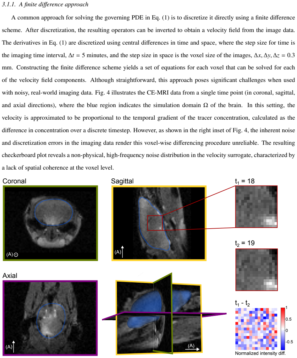
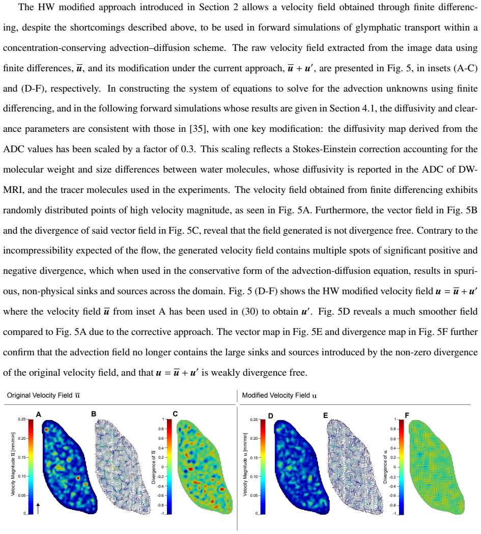
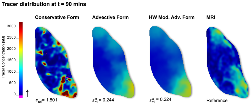
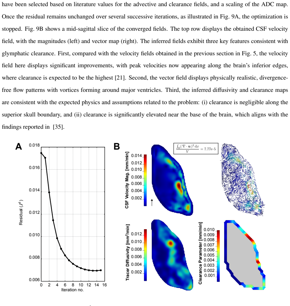
Recognition: unknown
Reconstruction of glymphatic transport fields from subject-specific imaging data, with particular emphasis on cerebrospinal fluid flow and tracer conservation
Pith reviewed 2026-05-09 18:16 UTC · model grok-4.3
The pith
An advection-diffusion model with velocity decomposition reconstructs divergence-free CSF transport fields from subject-specific brain imaging data.
A machine-rendered reading of the paper's core claim, the machinery that carries it, and where it could break.
Core claim
The authors show that an advection-diffusion model equipped with a velocity decomposition that imposes mass conservation permits the solution of a constrained inverse problem whose output is a set of spatially varying, solenoidal CSF velocity fields together with diffusivity and clearance parameters. Discretization via immersed isogeometric analysis with quadratic B-spline basis functions supplies the necessary smoothness and regularization. Forward simulations driven by the recovered fields reproduce the measured spatiotemporal tracer distributions, thereby confirming that the reconstructed transport quantities remain consistent with the underlying physics of incompressible flow and tracer,
What carries the argument
Advection-diffusion model with velocity decomposition enforcing zero divergence, solved as a constrained inverse problem and discretized by immersed isogeometric analysis with quadratic B-splines.
If this is right
- Spatially varying estimates of CSF velocity, diffusivity, and clearance parameters are obtained directly from contrast-enhanced MRI data.
- Forward simulations driven by the reconstructed fields reproduce experimental tracer observations with close quantitative agreement.
- The recovered velocity fields remain divergence-free, thereby preserving mass conservation throughout the transport domain.
- The framework supplies a generalizable route to infer physically consistent transport fields from noisy, imperfect imaging data in biological flow systems.
Where Pith is reading between the lines
- The same conservation-enforcing reconstruction could be applied to human glymphatic imaging to produce patient-specific clearance maps for neurodegenerative disease studies.
- Extension to other advection-dominated transport problems in porous media or vascular imaging would follow directly from the velocity-decomposition step.
- Validation against synthetic data with known ground-truth flows would quantify how measurement noise propagates into the recovered parameters.
- Coupling the method to multi-tracer experiments could separate advective from diffusive contributions at finer spatial scales.
Load-bearing premise
The advection-diffusion equation together with the proposed velocity decomposition is assumed to capture the essential physics of glymphatic transport, and the constrained inverse problem is assumed to admit unique, physically admissible solutions despite imaging noise and model error.
What would settle it
Forward simulation of tracer transport using the recovered velocity, diffusivity, and clearance fields would fail to reproduce the original spatiotemporal imaging data within measurement uncertainty, or the reconstructed velocity fields would display significant nonzero divergence.
Figures


read the original abstract
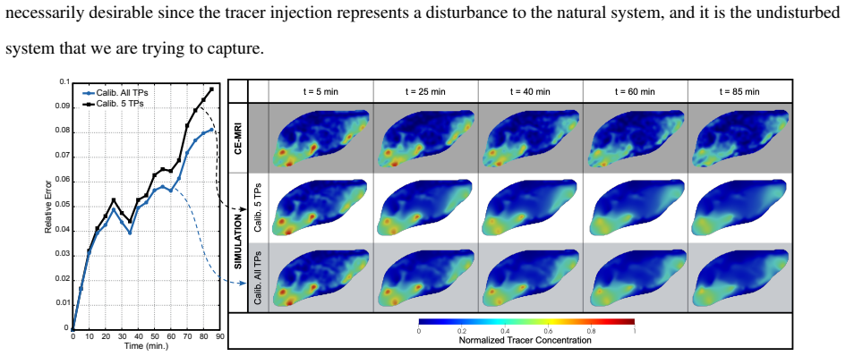
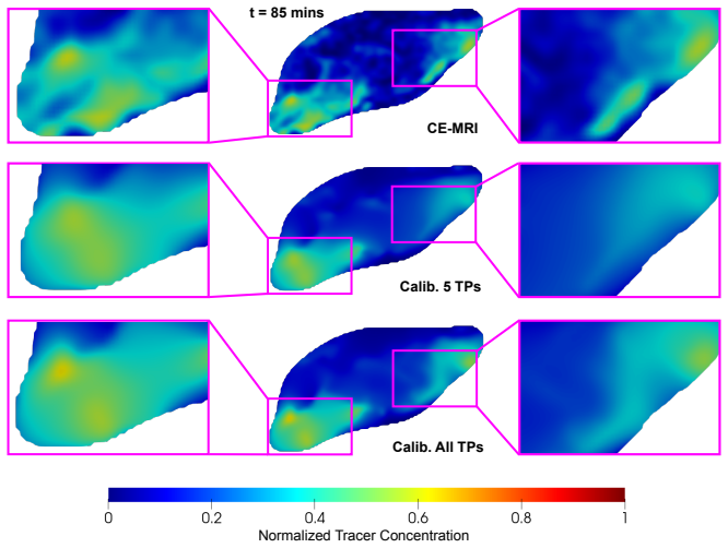
The reconstruction of physically valid transport fields from subject-specific imaging data is a fundamental challenge in image-based computational modeling due to measurement noise, modeling uncertainties and discretization errors. Without a methodology to construct models that faithfully reflect the underlying physics, mechanistic understanding of complex biological systems is inherently limited. In this work, we address this challenge in the glymphatic system, the brain's waste-clearance network, where cerebrospinal fluid (CSF) is transported through perivascular spaces into the brain parenchyma to facilitate metabolic waste removal. We introduce a computational framework for the high-fidelity reconstruction of subject-specific glymphatic transport fields from spatiotemporal imaging data. The formulation utilizes an advection-diffusion model with a velocity decomposition that imposes mass conservation, enabling the recovery of solenoidal (divergence-free) velocity fields through the solution of a constrained inverse problem. The system is discretized using immersed isogeometric analysis with quadratic B-spline basis functions, providing smooth, high-continuity solutions and inherent regularization of imaging noise. We demonstrate the framework's utility by using contrast-enhanced magnetic resonance imaging of tracer transport in a mouse brain, obtaining spatially varying estimates of CSF velocity, diffusivity, and clearance parameters. Forward simulations using the recovered fields show close agreement with experimental observations, validating the framework's ability to characterize complex transport dynamics while preserving physical integrity. This approach provides a generalizable methodology for the robust inference of physically consistent transport fields from imperfect imaging data, with broad applicability to the image-guided modeling of biological and engineering systems.
Editorial analysis
A structured set of objections, weighed in public.
Referee Report
Summary. The manuscript presents a computational framework for reconstructing subject-specific glymphatic transport fields (CSF velocity, diffusivity, and clearance parameters) from contrast-enhanced MRI tracer data in a mouse brain. It employs an advection-diffusion model with a velocity decomposition that enforces mass conservation by construction (yielding solenoidal, divergence-free velocity fields) solved as a constrained inverse problem, discretized via immersed isogeometric analysis using quadratic B-spline basis functions for smoothness and noise regularization. Forward simulations with the recovered fields are reported to show close agreement with experimental observations, validating the approach for physically consistent transport modeling.
Significance. If the central claims hold, this provides a useful methodology for image-based inference of physically valid advection-diffusion fields in biological systems, with built-in conservation properties and regularization suited to noisy data. The IGA discretization and velocity decomposition are strengths that could generalize beyond glymphatics to other transport inverse problems in computational biology and engineering.
major comments (2)
- [Abstract] Abstract: the validation statement that 'forward simulations using the recovered fields show close agreement with experimental observations, validating the framework' relies on agreement with the same fitted data and does not test uniqueness or robustness of the inverse recovery. No synthetic-data experiments with known ground-truth velocity, diffusivity, and clearance fields (or stability/condition-number analysis) are described to address the classical ill-posedness of advection-diffusion velocity inversion, even with the solenoidal constraint and B-spline regularization; this is load-bearing for the claim of recovering unique, physically valid fields.
- [Formulation] Formulation section (velocity decomposition and constrained inverse problem): while the decomposition imposing div v = 0 is a positive feature for conservation, the manuscript provides no quantitative recovery-error metrics, noise-sensitivity tests, or uniqueness guarantees on controlled data, leaving open whether multiple distinct solenoidal fields could fit the observations within imaging noise.
minor comments (2)
- [Abstract] Abstract and results: quantitative metrics (e.g., L2 norms, correlation coefficients, or error bars) for the reported 'close agreement' between forward simulations and observations are not provided, nor are details on data exclusion, noise handling, or parameter initialization.
- [Discretization] The manuscript would benefit from explicit discussion of how the quadratic B-spline continuity order and immersed boundary treatment interact with the regularization of the inverse problem.
Simulated Author's Rebuttal
We thank the referee for the constructive feedback and for recognizing the strengths of the velocity decomposition and IGA discretization. We address each major comment below and will revise the manuscript to incorporate additional validation analyses.
read point-by-point responses
-
Referee: [Abstract] Abstract: the validation statement that 'forward simulations using the recovered fields show close agreement with experimental observations, validating the framework' relies on agreement with the same fitted data and does not test uniqueness or robustness of the inverse recovery. No synthetic-data experiments with known ground-truth velocity, diffusivity, and clearance fields (or stability/condition-number analysis) are described to address the classical ill-posedness of advection-diffusion velocity inversion, even with the solenoidal constraint and B-spline regularization; this is load-bearing for the claim of recovering unique, physically valid fields.
Authors: We agree that agreement with the fitted experimental data alone does not establish uniqueness or robustness against the ill-posedness of the inverse problem. In the revised manuscript we will add a dedicated section presenting synthetic-data experiments. These will prescribe known ground-truth solenoidal velocity, diffusivity, and clearance fields, add controlled noise levels representative of imaging data, recover the fields via the same constrained inverse problem, and report quantitative recovery errors (L2 norms and relative errors). We will also include a condition-number analysis of the discretized system under the solenoidal constraint and quadratic B-spline regularization to quantify stability. revision: yes
-
Referee: [Formulation] Formulation section (velocity decomposition and constrained inverse problem): while the decomposition imposing div v = 0 is a positive feature for conservation, the manuscript provides no quantitative recovery-error metrics, noise-sensitivity tests, or uniqueness guarantees on controlled data, leaving open whether multiple distinct solenoidal fields could fit the observations within imaging noise.
Authors: We acknowledge that the current manuscript lacks quantitative recovery-error metrics and noise-sensitivity tests on controlled data. As described in the response to the abstract comment, the added synthetic experiments will supply these metrics (recovery errors versus ground truth and across noise levels) together with noise-sensitivity results. On uniqueness, we note that the solenoidal constraint reduces the admissible function space and the quadratic B-spline regularization further limits non-uniqueness in practice; however, a rigorous theoretical uniqueness proof for the continuous inverse problem is not available and would require additional assumptions on the data. The synthetic tests will instead demonstrate that, for the noise levels and regularization parameters used, the recovered fields remain close to the prescribed ground truth, thereby providing practical evidence that distinct solenoidal fields do not fit the observations equally well. revision: yes
Circularity Check
No significant circularity in the inverse reconstruction framework
full rationale
The paper recovers subject-specific velocity, diffusivity, and clearance fields by solving a constrained inverse problem whose inputs are external contrast-enhanced MRI tracer data. The advection-diffusion model with velocity decomposition enforces divergence-free velocity by construction, but the particular field values are determined by the data fit rather than by redefinition or tautology. No self-citations, uniqueness theorems imported from prior author work, or fitted quantities relabeled as independent predictions appear in the derivation. Forward simulation agreement with the same observations is standard inverse-problem validation and does not reduce the central claim to an input by construction. The chain is therefore self-contained against the external imaging benchmark.
Axiom & Free-Parameter Ledger
free parameters (2)
- spatially varying diffusivity
- spatially varying clearance parameters
axioms (3)
- domain assumption Glymphatic transport follows an advection-diffusion equation
- domain assumption Velocity field can be decomposed to enforce divergence-free condition
- standard math Immersed isogeometric analysis with quadratic B-splines provides suitable discretization and regularization
Reference graph
Works this paper leans on
-
[1]
S. S. Hossain, S. F. A. Hossainy, Y . Bazilevs, V . M. Calo, T. J. R. Hughes, Mathematical modeling of cou- pled drug and drug-encapsulated nanoparticle transport in patient-specific coronary artery walls, Computational Mechanics 49 (2) (2012) 213–242.doi:10.1007/s00466-011-0633-2
-
[2]
S. S. Hossain, Y . Zhang, X. Liang, F. Hussain, M. Ferrari, T. J. Hughes, P. Decuzzi, In Silico Vas- cular Modeling for Personalized Nanoparticle Delivery, Nanomedicine 8 (3) (2013) 343–357, _eprint: https://doi.org/10.2217/nnm.12.124.doi:10.2217/nnm.12.124. URLhttps://doi.org/10.2217/nnm.12.124
work page doi:10.2217/nnm.12.124.doi:10.2217/nnm.12.124 2013
-
[3]
T. Bohr, P. G. Hjorth, S. C. Holst, S. Hrabetová, V . Kiviniemi, T. Lilius, I. Lundgaard, K.-A. Mardal, E. A. Martens, Y . Mori, U. V . Nägerl, C. Nicholson, A. Tannenbaum, J. H. Thomas, J. Tithof, H. Benveniste, J. J. Iliff, D. H. Kelley, M. Nedergaard, The glymphatic system: Current understanding and modeling, iScience 25 (9) (2022) 104987.doi:10.1016/j...
-
[4]
J. J. Iliff, M. Wang, Y . Liao, B. A. Plogg, W. Peng, G. A. Gundersen, H. Benveniste, G. E. Vates, R. Deane, S. A. Goldman, E. A. Nagelhus, M. Nedergaard, A Paravascular Pathway Facilitates CSF Flow Through the Brain Parenchyma and the Clearance of Interstitial Solutes, Including Amyloidβ, Science Translational Medicine 4 (147) (2012) 147ra111–147ra111, p...
-
[5]
H. Mestre, Y . Mori, M. Nedergaard, The Brain’s Glymphatic System: Current Controversies, Trends in Neuro- sciences 43 (7) (2020) 458–466.doi:10.1016/j.tins.2020.04.003. URLhttps://www.cell.com/trends/neurosciences/abstract/S0166-2236(20)30077-1
-
[6]
Y . Guo, K. Quirk, D. H. Kelley, J. H. Thomas, Advection and diffusion in perivascular and extracellular spaces in the brain, J R Soc Interface 22 (226) (2025) 20250010.doi:10.1098/rsif.2025.0010
-
[7]
L. M. Hablitz, M. Nedergaard, The Glymphatic System: A Novel Component of Fundamental Neurobiology, J. Neurosci. 41 (37) (2021) 7698–7711.doi:10.1523/JNEUROSCI.0619-21.2021. URLhttps://www.jneurosci.org/lookup/doi/10.1523/JNEUROSCI.0619-21.2021
-
[8]
N. MacAulay, Molecular mechanisms of brain water transport, Nat Rev Neurosci 22 (6) (2021) 326–344, pub- lisher: Nature Publishing Group.doi:10.1038/s41583-021-00454-8. URLhttps://www.nature.com/articles/s41583-021-00454-8
-
[9]
F. R. Buccellato, M. D’Anca, M. Serpente, A. Arighi, D. Galimberti, The Role of Glymphatic System in Alzheimer’s and Parkinson’s Disease Pathogenesis, Biomedicines 10 (9) (2022) 2261, publisher: Multidisci- plinary Digital Publishing Institute.doi:10.3390/biomedicines10092261. URLhttps://www.mdpi.com/2227-9059/10/9/2261
-
[10]
S. B. Hladky, M. A. Barrand, The glymphatic hypothesis: the theory and the evidence, Fluids Barriers CNS 19 (1) (2022) 9.doi:10.1186/s12987-021-00282-z. URLhttps://doi.org/10.1186/s12987-021-00282-z
-
[11]
T. Vikner, K. M. Johnson, R. V . Cadman, T. J. Betthauser, R. E. Wilson, N. Chin, L. B. Eisenmenger, S. C. Johnson, L. A. Rivera-Rivera, CSF dynamics throughout the ventricular system using 4D flow MRI: associations to arterial pulsatility, ventricular volumes, and age, Fluids Barriers CNS 21 (1) (2024) 68.doi:10.1186/ s12987-024-00570-4. URLhttps://doi.o...
-
[12]
J. J. Iliff, H. Lee, M. Yu, T. Feng, J. Logan, M. Nedergaard, H. Benveniste, Brain-wide pathway for waste clear- ance captured by contrast-enhanced MRI, J. Clin. Invest. 123 (3) (2013) 1299–1309.doi:10.1172/JCI67677. URLhttp://www.jci.org/articles/view/67677 37
-
[13]
D. S. Lee, M. Suh, A. Sarker, Y . Choi, Brain Glymphatic/Lymphatic Imaging by MRI and PET, Nucl Med Mol Imaging 54 (5) (2020) 207–223.doi:10.1007/s13139-020-00665-4. URLhttps://doi.org/10.1007/s13139-020-00665-4
-
[14]
Y . Wu, F. Xu, D. Zhu, A. M. Li, K. Wang, Q. Qin, J. Xu, Cerebrospinal fluid flow within ventricles and subarachnoid space evaluated by velocity selective spin labeling MRI, NeuroImage 309 (2025) 121095. doi:10.1016/j.neuroimage.2025.121095. URLhttps://linkinghub.elsevier.com/retrieve/pii/S1053811925000977
-
[15]
A. A. Badachhape, P. K. Working, M. Srivastava, P. Bhandari, I. V . Stupin, L. Devkota, E. A. Tanifum, A. V . Annapragada, K. B. Ghaghada, Pre-clinical dose-ranging efficacy, pharmacokinetics, tissue biodistribution, and toxicity of a targeted contrast agent for MRI of amyloid deposition in Alzheimer’s disease, Sci Rep 10 (1) (2020) 16185, publisher: Natu...
-
[16]
M. Asgari, D. de Zélicourt, V . Kurtcuoglu, Glymphatic solute transport does not require bulk flow, Sci Rep 6 (1) (2016) 38635.doi:10.1038/srep38635. URLhttps://www.nature.com/articles/srep38635
-
[17]
https://doi.org/https://doi.org/10.1016/j
A. Solheim, G. Ringstad, P. K. Eide, K.-A. Mardal, Geometry Reduced Order Modeling (GROM) with appli- cation to modeling of glymphatic function, Brain Research Bulletin 231 (2025) 111558.doi:10.1016/j. brainresbull.2025.111558. URLhttps://linkinghub.elsevier.com/retrieve/pii/S0361923025003703
work page doi:10.1016/j 2025
-
[18]
K. E. Holter, B. Kehlet, A. Devor, T. J. Sejnowski, A. M. Dale, S. W. Omholt, O. P. Ottersen, E. A. Nagelhus, K.-A. Mardal, K. H. Pettersen, Interstitial solute transport in 3D reconstructed neuropil occurs by diffusion rather than bulk flow, Proceedings of the National Academy of Sciences 114 (37) (2017) 9894–9899.doi: 10.1073/pnas.1706942114. URLhttps:/...
-
[19]
L. Ray, J. J. Iliff, J. J. Heys, Analysis of convective and diffusive transport in the brain interstitium, Fluids Barriers CNS 16 (1) (2019) 6.doi:10.1186/s12987-019-0126-9. URLhttps://doi.org/10.1186/s12987-019-0126-9
-
[20]
S. Koundal, R. Elkin, S. Nadeem, Y . Xue, S. Constantinou, S. Sanggaard, X. Liu, B. Monte, F. Xu, W. Van Nos- trand, M. Nedergaard, H. Lee, J. Wardlaw, H. Benveniste, A. Tannenbaum, Optimal Mass Transport with La- grangian Workflow Reveals Advective and Diffusion Driven Solute Transport in the Glymphatic System, Sci Rep 10 (1) (2020) 1990.doi:10.1038/s415...
-
[21]
H. Benveniste, H. Lee, B. Ozturk, X. Chen, S. Koundal, P. Vaska, A. Tannenbaum, N. D. V olkow, Glymphatic Cerebrospinal Fluid and Solute Transport Quantified by MRI and PET Imaging, Neuroscience 474 (2021) 63– 79.doi:10.1016/j.neuroscience.2020.11.014. URLhttps://linkinghub.elsevier.com/retrieve/pii/S0306452220307302
-
[22]
V . Vinje, B. Zapf, G. Ringstad, P. K. Eide, M. E. Rognes, K.-A. Mardal, Human brain solute transport quantified by glymphatic MRI-informed biophysics during sleep and sleep deprivation, Fluids Barriers CNS 20 (1) (2023) 62.doi:10.1186/s12987-023-00459-8. URLhttps://doi.org/10.1186/s12987-023-00459-8
-
[23]
X. Chen, H. Benveniste, A. R. Tannenbaum, Unbalanced regularized optimal mass transport with applications to fluid flows in the brain, Sci Rep 14 (1) (2024) 1111.doi:10.1038/s41598-023-50874-y. URLhttps://www.nature.com/articles/s41598-023-50874-y 38
-
[24]
X.-B. Ding, X.-X. Wang, D.-H. Xia, H. Liu, H.-Y . Tian, Y . Fu, Y .-K. Chen, C. Qin, J.-Q. Wang, Z. Xiang, Z.-X. Zhang, Q.-C. Cao, W. Wang, J.-Y . Li, E. Wu, B.-S. Tang, M.-M. Ma, J.-F. Teng, X.-J. Wang, Impaired meningeal lymphatic drainage in patients with idiopathic Parkinson’s disease, Nat Med 27 (3) (2021) 411–418, publisher: Nature Publishing Group....
-
[25]
Kope ´c, D
K. Kope ´c, D. Koziorowski, S. Szlufik, The Therapeutic Potential of Glymphatic System Activity to Reduce the Pathogenic Accumulation of Cytotoxic Proteins in Alzheimer’s Disease, International Journal of Molecu- lar Sciences 26 (15) (2025) 7552, publisher: Multidisciplinary Digital Publishing Institute.doi:10.3390/ ijms26157552. URLhttps://www.mdpi.com/1...
2025
-
[26]
A. D. Bakiler, Created in biorender, BioRender.com,https://BioRender.com/4y16xof(2026)
2026
-
[27]
M. Loecher, S. Kecskemeti, P. Turski, O. Wieben, Comparison of divergence-free algorithms for 3D MRI with three-directional velocity encoding, J Cardiovasc Magn Reson 14 (1) (2012) W64.doi:10.1186/ 1532-429X-14-S1-W64. URLhttps://doi.org/10.1186/1532-429X-14-S1-W64
-
[28]
S. M. Song, S. Napel, G. H. Glover, N. J. Pelc, Noise reduction in three-dimensional phase-contrast MR velocity measurementsl, Journal of Magnetic Resonance Imaging 3 (4) (1993) 587–596, _eprint: https://onlinelibrary.wiley.com/doi/pdf/10.1002/jmri.1880030407.doi:10.1002/jmri.1880030407. URLhttps://onlinelibrary.wiley.com/doi/abs/10.1002/jmri.1880030407
work page doi:10.1002/jmri.1880030407.doi:10.1002/jmri.1880030407 1993
-
[29]
J. Busch, D. Giese, L. Wissmann, S. Kozerke, Reconstruction of divergence-free velocity fields from cine 3D phase-contrast flow measurements, Magnetic Resonance in Medicine 69 (1) (2013) 200–210, _eprint: https://onlinelibrary.wiley.com/doi/pdf/10.1002/mrm.24221.doi:10.1002/mrm.24221. URLhttps://onlinelibrary.wiley.com/doi/abs/10.1002/mrm.24221
-
[30]
F. Ong, M. Uecker, U. Tariq, A. Hsiao, M. T. Alley, S. S. Vasanawala, M. Lustig, Robust 4D flow denoising using divergence-free wavelet transform, Magnetic Resonance in Medicine 73 (2) (2015) 828–842, _eprint: https://onlinelibrary.wiley.com/doi/pdf/10.1002/mrm.25176.doi:10.1002/mrm.25176. URLhttps://onlinelibrary.wiley.com/doi/abs/10.1002/mrm.25176
-
[31]
H. Mestre, J. Tithof, T. Du, W. Song, W. Peng, A. M. Sweeney, G. Olveda, J. H. Thomas, M. Nedergaard, D. H. Kelley, Flow of cerebrospinal fluid is driven by arterial pulsations and is reduced in hypertension, Nat Commun 9 (1) (2018) 4878, publisher: Nature Publishing Group.doi:10.1038/s41467-018-07318-3. URLhttps://www.nature.com/articles/s41467-018-07318-3
-
[32]
G. Meng, J. Zhong, Q. Zhang, J. S. J. Wong, J. Wu, K. K. Tsia, N. Ji, Ultrafast two-photon fluorescence imaging of cerebral blood circulation in the mouse brain in vivo, Proceedings of the National Academy of Sciences 119 (23) (2022) e2117346119.doi:10.1073/pnas.2117346119. URLhttps://www.pnas.org/doi/full/10.1073/pnas.2117346119
-
[33]
T. J. Hughes, G. N. Wells, Conservation properties for the Galerkin and stabilised forms of the advec- tion–diffusion and incompressible Navier–Stokes equations, Computer Methods in Applied Mechanics and En- gineering 194 (9-11) (2005) 1141–1159.doi:10.1016/j.cma.2004.06.034. URLhttps://linkinghub.elsevier.com/retrieve/pii/S0045782504003172
-
[34]
A. N. Brooks, T. J. R. Hughes, Streamline upwind/Petrov-Galerkin formulations for convection dominated flows with particular emphasis on the incompressible Navier-Stokes equations, Computer Methods in Applied Me- chanics and Engineering 32 (1) (1982) 199–259.doi:10.1016/0045-7825(82)90071-8. URLhttps://www.sciencedirect.com/science/article/pii/0045782582900718 39
-
[35]
M. J. Johnson, M. R. Abdelmalik, F. A. Baidoo, A. Badachhape, T. J. Hughes, S. S. Hossain, Image-guided subject-specific modeling of glymphatic transport and amyloid deposition, Computer Methods in Applied Me- chanics and Engineering 417 (2023) 116449.doi:10.1016/j.cma.2023.116449. URLhttps://linkinghub.elsevier.com/retrieve/pii/S004578252300573X
-
[36]
A. Düster, J. Parvizian, Z. Yang, E. Rank, The finite cell method for three-dimensional problems of solid me- chanics, Computer Methods in Applied Mechanics and Engineering 197 (45-48) (2008) 3768–3782.doi: 10.1016/j.cma.2008.02.036. URLhttps://linkinghub.elsevier.com/retrieve/pii/S0045782508001163
-
[37]
C. Giannelli, B. Jüttler, S. K. Kleiss, A. Mantzaflaris, B. Simeon, J.Speh, THB-splines: An effective mathe- matical technology for adaptive refinement in geometric design and isogeometric analysis, Computer Methods in Applied Mechanics and Engineering 299 (2016) 337–365.doi:10.1016/j.cma.2015.11.002. URLhttps://linkinghub.elsevier.com/retrieve/pii/S00457...
-
[38]
A. Buffa, C. Giannelli, P. Morgenstern, D. Peterseim, Complexity of hierarchical refinement for a class of ad- missible mesh configurations, Computer Aided Geometric Design 47 (2016) 83–92.doi:10.1016/j.cagd. 2016.04.003. URLhttps://linkinghub.elsevier.com/retrieve/pii/S0167839616300449
-
[39]
J. van Zwieten, G. van Zwieten, W. Hoitinga, Nutils 9.0 (2025).doi:10.5281/zenodo.6006701
-
[40]
A. J. Smith, X. Yao, J. A. Dix, B.-J. Jin, A. S. Verkman, Test of the ’glymphatic’ hypothesis demonstrates diffusive and aquaporin-4-independent solute transport in rodent brain parenchyma, eLife 6 (2017) e27679. doi:10.7554/eLife.27679. URLhttps://doi.org/10.7554/eLife.27679
-
[41]
A. Aspelund, S. Antila, S. T. Proulx, T. V . Karlsen, S. Karaman, M. Detmar, H. Wiig, K. Alitalo, A dural lymphatic vascular system that drains brain interstitial fluid and macromolecules, Journal of Experimental Medicine 212 (7) (2015) 991–999.doi:10.1084/jem.20142290. URLhttps://rupress.org/jem/article/212/7/991/41853/A-dural-lymphatic-vascular-system-t...
-
[42]
Q. Ma, B. V . Ineichen, M. Detmar, S. T. Proulx, Outflow of cerebrospinal fluid is predominantly through lymphatic vessels and is reduced in aged mice, Nat Commun 8 (1) (2017) 1434.doi:10.1038/ s41467-017-01484-6. URLhttps://www.nature.com/articles/s41467-017-01484-6
2017
-
[43]
L. Xie, H. Kang, Q. Xu, M. J. Chen, Y . Liao, M. Thiyagarajan, J. O’Donnell, D. J. Christensen, C. Nicholson, J. J. Iliff, T. Takano, R. Deane, M. Nedergaard, Sleep drives metabolite clearance from the adult brain, Science 342 (6156) (2013) 373–377.arXiv:https://www.science.org/doi/pdf/10.1126/science.1241224, doi:10.1126/science.1241224. URLhttps://www.s...
discussion (0)
Sign in with ORCID, Apple, or X to comment. Anyone can read and Pith papers without signing in.