Recognition: 1 theorem link
Approaching human parity in the quality of automated organoid image segmentation
Pith reviewed 2026-05-08 18:46 UTC · model grok-4.3
The pith
A composite method pairing the Segment Anything Model with a domain-specific tool segments organoid images at or near inter-observer human accuracy.
A machine-rendered reading of the paper's core claim, the machinery that carries it, and where it could break.
Core claim
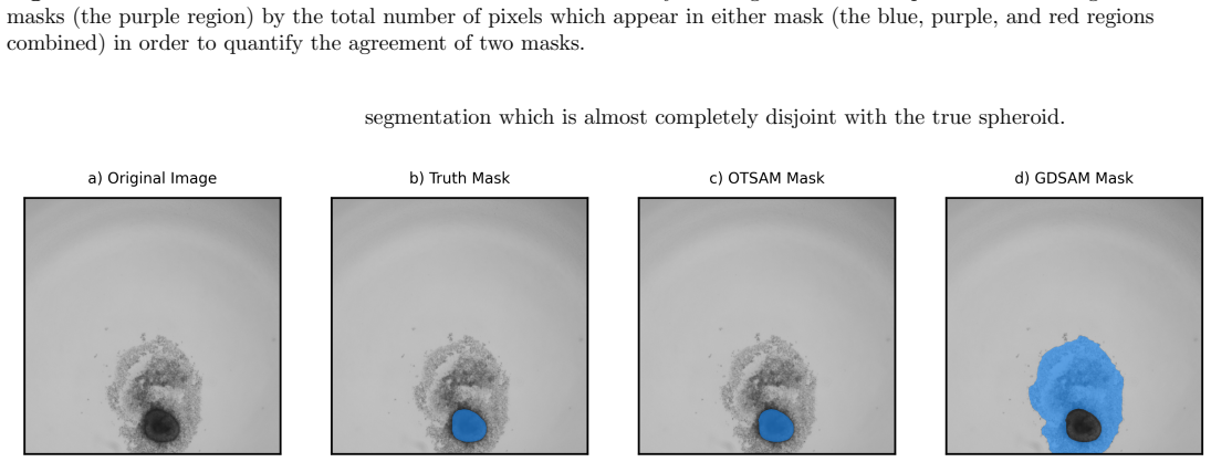
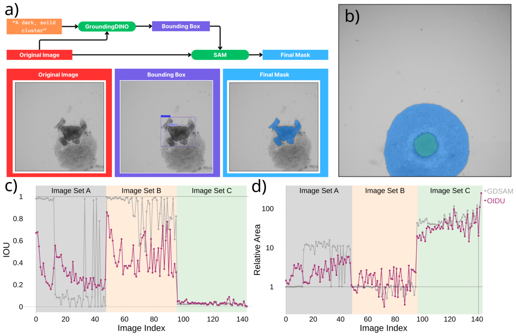
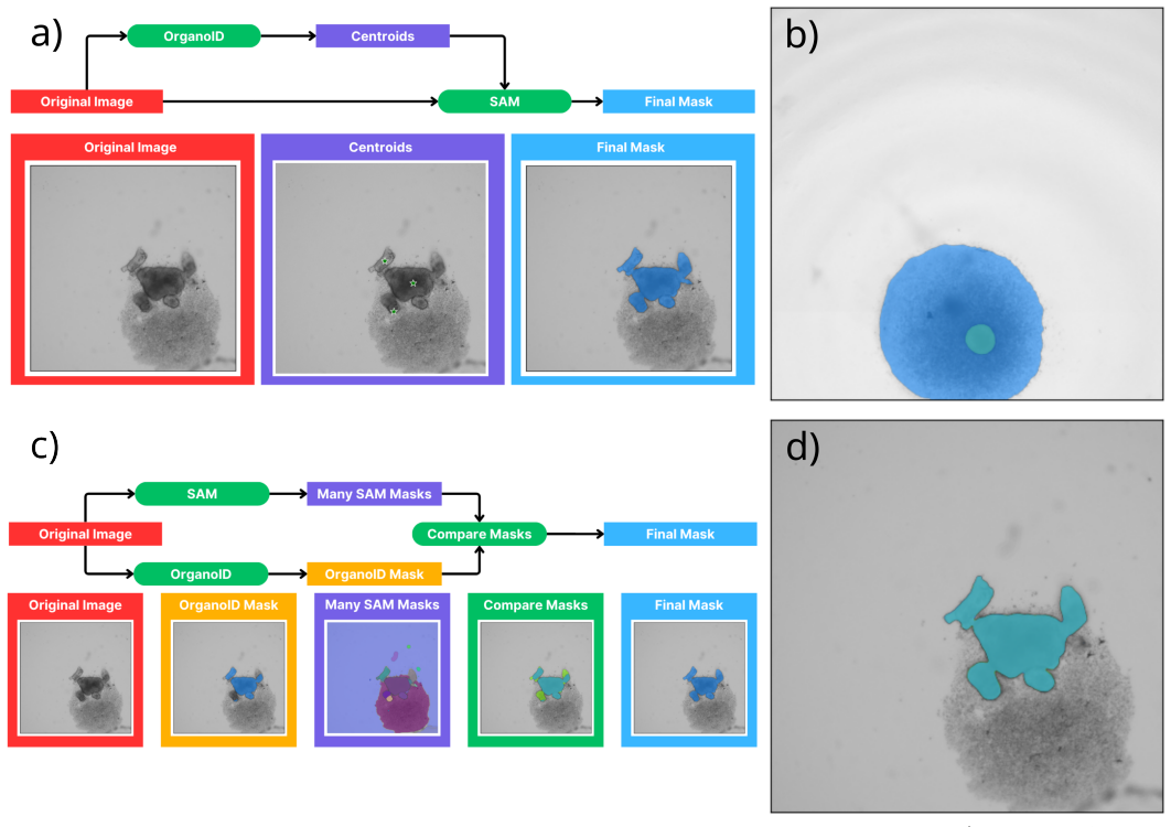
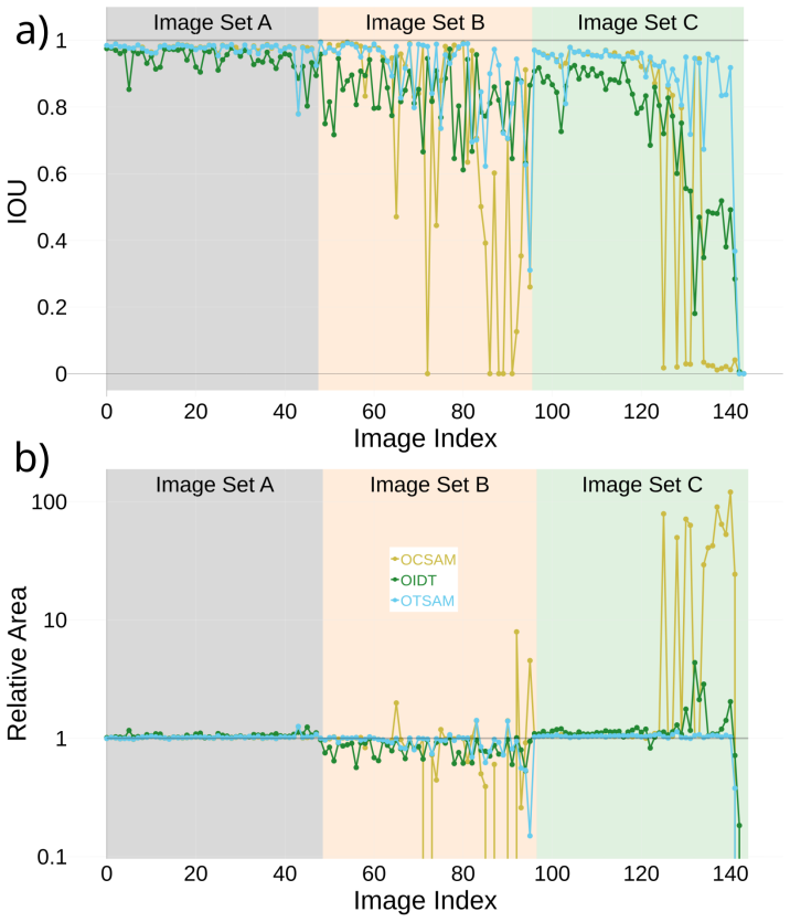
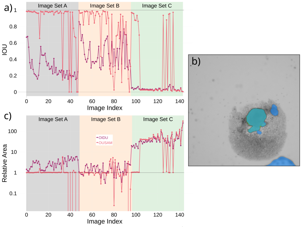
No single existing segmentation tool delivers sufficient accuracy on every test image of pluripotent-stem-cell-derived spheroids, yet the composite method that combines the Segment Anything Model with a domain-specific tool produces consistent and accurate results on all but a very small fraction of the most challenging images; by one quantitative measure its performance equals inter-observer variability among human annotators and by others it lies very close to that benchmark.
What carries the argument
The composite segmentation pipeline that applies the Segment Anything Model (SAM) as a general-purpose foundation model and then refines its output with an existing domain-specific organoid segmentation tool.
If this is right
- Large-scale time-lapse studies of organoid development can replace most manual outlining with automated measurements.
- Morphological changes during disease modeling become easier to quantify across hundreds of organoids.
- The same hybrid strategy can be tested on other complex three-dimensional cell cultures where single tools currently fall short.
- Routine monitoring of organoid size and shape no longer requires an expert annotator for every image.
Where Pith is reading between the lines
- The same SAM-plus-refinement pattern may transfer to other biomedical imaging domains that already possess one reliable but narrow tool.
- If the composite method maintains its performance on live-cell imaging sequences, it could enable fully automated tracking of organoid growth trajectories.
- Adoption would shift the bottleneck in organoid research from image segmentation to downstream biological interpretation of the resulting shape and size data.
Load-bearing premise
The selected test images and the manual annotations used as ground truth adequately represent the full range of real-world organoid imaging conditions and that matching inter-observer variability is the appropriate target for acceptable automated performance.
What would settle it
Apply the composite method to a new collection of organoid images drawn from different laboratories, imaging modalities, or organoid types and measure whether its segmentation error exceeds the inter-observer variability measured on the same set.
Figures


read the original abstract
Organoids are complex, three dimensional, self-organizing cell cultures which manifest organ-like features and represent a powerful platform for studying human disease and developing treatment options. Organoid development is characterized by dynamic morphological and cellular organization, which mimic some aspects of organ development. To study these rapid changes over the course of organoid development, advanced imaging and analytical tools are critical to accurately monitor the trajectory of organoid growth and investigate disease processes. In this work, we focus on computer vision and machine learning techniques to automatically measure the size and shape of developing spheroids derived from pluripotent stem cells (iPSCs), which are typically the starting material for generating organoid cultures. To facilitate this task, we introduce a composite method that combines the Segment Anything Model (SAM), a general-purpose foundation model, with an existing domain-specific tool. This composite method is evaluated together with several existing tools by testing them on organoid image data and comparing with the results of manual image segmentation. We find that no single existing tool is able to segment the test images with sufficient accuracy across all test conditions, but the newly introduced composite method produces consistent and accurate results for all but a very small fraction of the most challenging images. Finally, we compare the accuracy of this method to the variability between manual segmentations by independent annotators (inter-observer variability) and find that by one measure it performs at the level of inter-observer variability and by others it performs very close to it.
Editorial analysis
A structured set of objections, weighed in public.
Referee Report
Summary. The manuscript introduces a composite segmentation approach that combines the Segment Anything Model (SAM) with an existing domain-specific tool for automated analysis of organoid images derived from iPSCs. It evaluates the method against several existing tools and manual segmentations, claiming consistent and accurate results for all but a small fraction of challenging images, with performance reaching inter-observer variability on one metric and approaching it on others.
Significance. If the quantitative results hold with representative data, the work could enable scalable, reproducible monitoring of organoid morphology in developmental biology and disease modeling, reducing dependence on manual annotation. The direct comparison to inter-observer variability and use of a foundation model adapted to the domain are notable strengths that support practical utility.
major comments (2)
- Abstract: The central claim that the composite method 'performs at the level of inter-observer variability' by one measure and 'very close to it' by others is not supported by any reported numerical values for the metrics (Dice, IoU, boundary error, etc.), dataset cardinality, image selection protocol, or characterization of the 'very small fraction' of failure cases. These details are load-bearing for evaluating statistical robustness and external validity of the human-parity assertion.
- Evaluation section: The manuscript adopts inter-observer variability as the benchmark for acceptable automated performance without explicit justification or analysis of whether this reference distribution aligns with biological requirements (e.g., tolerance for error in downstream growth trajectory studies); this assumption requires concrete support to sustain the parity conclusion.
minor comments (2)
- Clarify the precise integration mechanism between SAM and the domain-specific tool, including any post-processing steps or parameter choices, to support reproducibility.
- Add representative failure-case images and quantitative breakdowns by image difficulty or morphology type to the results or supplementary material.
Simulated Author's Rebuttal
We thank the referee for their detailed and constructive review of our manuscript. We address the major comments point by point below, proposing revisions to enhance the clarity and support for our claims.
read point-by-point responses
-
Referee: Abstract: The central claim that the composite method 'performs at the level of inter-observer variability' by one measure and 'very close to it' by others is not supported by any reported numerical values for the metrics (Dice, IoU, boundary error, etc.), dataset cardinality, image selection protocol, or characterization of the 'very small fraction' of failure cases. These details are load-bearing for evaluating statistical robustness and external validity of the human-parity assertion.
Authors: We acknowledge that the abstract would be improved by including specific supporting details. The manuscript does report the numerical values for the metrics, the dataset cardinality and selection protocol, and the characterization of failure cases in the Evaluation and Results sections. To make these load-bearing details immediately available to readers, we will revise the abstract to include key quantitative results and a summary of the dataset and failure cases. revision: yes
-
Referee: Evaluation section: The manuscript adopts inter-observer variability as the benchmark for acceptable automated performance without explicit justification or analysis of whether this reference distribution aligns with biological requirements (e.g., tolerance for error in downstream growth trajectory studies); this assumption requires concrete support to sustain the parity conclusion.
Authors: The use of inter-observer variability as a benchmark is standard practice in the field for assessing automated segmentation performance against human experts. However, we agree that explicit justification and analysis of its alignment with biological requirements would strengthen the manuscript. We will add a new paragraph in the Evaluation section that justifies this choice with references to prior work and discusses its implications for downstream applications such as growth trajectory studies. revision: yes
Circularity Check
No circularity: evaluation uses independent external manual annotations and inter-observer benchmarks
full rationale
The paper introduces a composite SAM-based segmentation method and evaluates it empirically on organoid images by direct comparison to manual segmentations performed by independent annotators. Performance is reported relative to inter-observer variability as an external reference. No equations, parameter fitting, or derivations are described that reduce the reported accuracy to the method's own inputs by construction. No self-citations are invoked as load-bearing uniqueness theorems or ansatzes. The central claim rests on external test data and human annotations rather than any self-referential loop, satisfying the criteria for a self-contained empirical result.
Axiom & Free-Parameter Ledger
axioms (1)
- domain assumption Manual segmentations by independent annotators constitute a reliable reference standard for measuring automated accuracy.
Reference graph
Works this paper leans on
-
[1]
Modeling Development and Disease with Organoids
Clevers H. Modeling Development and Disease with Organoids. Cell. 2016;165(7):1586–1597. doi:10.1016/j.cell.2016.05.082
-
[2]
Organoids: Modeling Development and the Stem Cell Niche in a Dish
Kretzschmar K, Clevers H. Organoids: Modeling Development and the Stem Cell Niche in a Dish. Developmental Cell. 2016;38(6):590–600. doi:10.1016/j.devcel.2016.08.014
-
[3]
Organoids — Preclinical Models of Human Disease
Li M, Belmonte JCI. Organoids — Preclinical Models of Human Disease. New England Journal of Medicine. 2019;380(6):569–579. doi:10.1056/NEJMra1806175
-
[5]
Hofer M, Lutolf MP. Engineering organoids. Nature Reviews Materials. 2021;6(5):402–420. doi:10.1038/s41578-021-00279-y
-
[6]
Importance of Organoids for Personalized Medicine
Perkhofer L, F Pierre-Olivier, M Martin, , Kleger A. Importance of Organoids for Personalized Medicine. Personalized Medicine. 2018;15(6):461–465. doi:10.2217/pme-2018-0071
-
[7]
Oral Mucosal Organoids as a Potential Platform for Personalized Cancer Therapy
Driehuis E, Kolders S, Spelier S, L˜ ohmussaar K, Willems SM, Devriese LA, et al. Oral Mucosal Organoids as a Potential Platform for Personalized Cancer Therapy. Cancer Discovery. 2019;9(7):852–871. doi:10.1158/2159-8290.CD-18-1522
-
[8]
Organoid based personalized medicine: from bench to bedside
Li Y, Tang P, Cai S, Peng J, Hua G. Organoid based personalized medicine: from bench to bedside. Cell Regeneration. 2020;9(1):21. doi:10.1186/s13619-020-00059-z
-
[9]
Organoid-based personalized medicine: from tumor outcome prediction to autologous transplantation
Soto-Gamez A, Gunawan JP, Barazzuol L, Pringle S, Coppes RP. Organoid-based personalized medicine: from tumor outcome prediction to autologous transplantation. Stem Cells. 2024;42(6):499–508. doi:10.1093/stmcls/sxae023
-
[10]
Patient-Derived Organoids: A Game-Changer in Personalized Cancer Medicine
Abbasian MH, Sobhani N, Sisakht MM, D’Angelo A, Sirico M, Roudi R. Patient-Derived Organoids: A Game-Changer in Personalized Cancer Medicine. Stem Cell Reviews and Reports. 2025;21(1):211–225. doi:10.1007/s12015-024-10805-4
-
[11]
Sarker IH. Deep Learning: A Comprehensive Overview on Techniques, Taxonomy, Applications and Research Directions. SN Computer Science. 2021;2(6):1–20. doi:10.1007/s42979-021-00815-1. May 6, 2026 24/26
-
[12]
A review on deep learning in medical image analysis
Suganyadevi S, Seethalakshmi V, Balasamy K. A review on deep learning in medical image analysis. International Journal of Multimedia Information Retrieval. 2021;11(1):19–38. doi:10.1007/s13735-021-00218-1
-
[13]
Deep Learning Applications in Medical Image Analysis
Ker J, Wang L, Rao J, Lim T. Deep Learning Applications in Medical Image Analysis. IEEE Access. 2018;6:9375–9389. doi:10.1109/ACCESS.2017.2788044
-
[14]
Deep learning for cellular image analysis
Moen E, Bannon D, Kudo T, Graf W, Covert M, Van Valen D. Deep learning for cellular image analysis. Nature Methods. 2019;16(12):1233–1246. doi:10.1038/s41592-019-0403-1
-
[15]
Frontiers| CNN-Based Cell Analysis: From Image to Quantitative Representation
Allier C, Herv´ e L, Paviolo C, Mandula O, Cioni O, Pierr´ e W, et al. Frontiers| CNN-Based Cell Analysis: From Image to Quantitative Representation. Frontiers in Physics. 2022;doi:10.3389/fphy.2021.776805
-
[16]
Growth of Epithelial Organoids in a Defined Hydrogel
Broguiere N, Isenmann L, Hirt C, Ringel T, Placzek S, Cavalli E, et al. Growth of Epithelial Organoids in a Defined Hydrogel. Advanced Materials. 2018;30(43):1801621. doi:10.1002/adma.201801621
-
[17]
MOrgAna: accessible quantitative analysis of organoids with machine learning
Gritti N, Lim JL, Anla¸ s K, Pandya M, Aalderink G, Mart´ ınez-Ara G, et al. MOrgAna: accessible quantitative analysis of organoids with machine learning. Development. 2021;148(18):dev199611. doi:10.1242/dev.199611
-
[18]
Development of a deep learning based image processing tool for enhanced organoid analysis
Park T, Kim TK, Han YD, Kim KA, Kim H, Kim HS. Development of a deep learning based image processing tool for enhanced organoid analysis. Scientific Reports. 2023;13(1):19841. doi:10.1038/s41598-023-46485-2
-
[19]
OrganoID: A versatile deep learning platform for tracking and analysis of single-organoid dynamics
Matthews JM, Schuster B, Kashaf SS, Liu P, Ben-Yishay R, Ishay-Ronen D, et al. OrganoID: A versatile deep learning platform for tracking and analysis of single-organoid dynamics. PLOS Computational Biology. 2022;18(11):e1010584. doi:10.1371/journal.pcbi.1010584
-
[20]
U-Net: Convolutional Networks for Biomedical Image Segmentation
Ronneberger O, Fischer P, Brox T. U-Net: Convolutional Networks for Biomedical Image Segmentation. In: Navab N, Hornegger J, Wells WM, Frangi AF, editors. Medical Image Computing and Computer-Assisted Intervention – MICCAI 2015. Munich, Germany: Springer International Publishing; 2015. p. 234–241. Available from:http://arxiv.org/abs/1505.04597
work page internal anchor Pith review arXiv 2015
-
[21]
MSU-Net: Multi-Scale U-Net for 2D Medical Image Segmentation
Su R, Zhang D, Liu J, Cheng C. MSU-Net: Multi-Scale U-Net for 2D Medical Image Segmentation. Frontiers in Genetics. 2021;doi:10.3389/fgene.2021.639930
-
[22]
Kirillov A, Mintun E, Ravi N, Mao H, Rolland C, Gustafson L, et al. Segment Anything. Preprint arXiv:230402643. 2023;doi:10.48550/arXiv.2304.02643
work page internal anchor Pith review doi:10.48550/arxiv.2304.02643 2023
-
[23]
Segment Anything for Microscopy
Archit A, Freckmann L, Nair S, Khalid N, Hilt P, Rajashekar V, et al. Segment Anything for Microscopy. Nature Methods. 2025;22(3):579–591. doi:10.1038/s41592-024-02580-4
-
[24]
Grounding DINO: Marrying DINO with Grounded Pre-Training for Open-Set Object Detection
Liu S, Zeng Z, Ren T, Li F, Zhang H, Yang J, et al.. Grounding DINO: Marrying DINO with Grounded Pre-Training for Open-Set Object Detection
-
[25]
Available from:http://arxiv.org/abs/2303.05499
-
[26]
Grounded SAM: Assembling Open-World Models for Diverse Visual Tasks
Ren T, Liu S, Zeng A, Lin J, Li K, Cao H, et al.. Grounded SAM: Assembling Open-World Models for Diverse Visual Tasks; 2024. Available from: http://arxiv.org/abs/2401.14159. May 6, 2026 25/26
work page Pith review arXiv 2024
-
[27]
Mumuni F, Mumuni A. Segment Anything Model for automated image data annotation: empirical studies using text prompts from Grounding DINO; 2024. Available from:http://arxiv.org/abs/2406.19057
-
[28]
Automated microfluidic platform for dynamic and combinatorial drug screening of tumor organoids
Schuster B, Junkin M, Kashaf SS, Romero-Calvo I, Kirby K, Matthews J, et al. Automated microfluidic platform for dynamic and combinatorial drug screening of tumor organoids. Nature Communications. 2020;11(1):5271. doi:10.1038/s41467-020-19058-4
-
[29]
Chen Y, Tristan CA, Chen L, Jovanovic VM, Malley C, Chu PH, et al. A versatile polypharmacology platform promotes cytoprotection and viability of human pluripotent and differentiated cells. Nat Methods. 2021;18(5):528–541. doi:10.1038/s41592-021-01126-2
-
[30]
scikit-image: image processing in Python,
Van Der Walt S, Sch¨ onberger JL, Nunez-Iglesias J, Boulogne F, Warner JD, Yager N, et al. scikit-image: image processing in Python. PeerJ. 2014;2:e453. doi:10.7717/peerj.453. May 6, 2026 26/26
discussion (0)
Sign in with ORCID, Apple, or X to comment. Anyone can read and Pith papers without signing in.