ResNet-34 with Lightweight Decoder for Accurate and Efficient Segmentation of Fetal Brain MRI
Pith reviewed 2026-06-28 16:20 UTC · model grok-4.3
The pith
ResNet-34 encoder paired with a lightweight MLP decoder segments fetal brain MRI more accurately than UNet variants while using fewer parameters.
A machine-rendered reading of the paper's core claim, the machinery that carries it, and where it could break.
Core claim
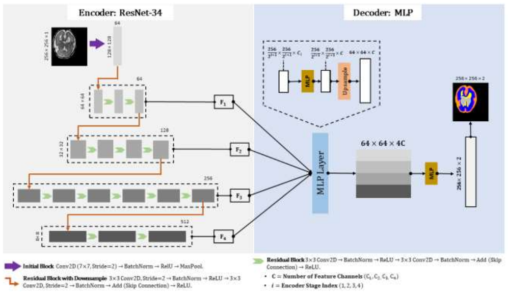
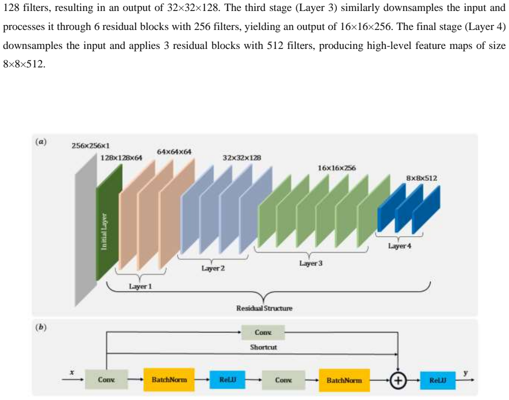
The model combines a ResNet-34 encoder with a lightweight decoder that incorporates MLP modules for adaptive feature refinement, preserves anatomical boundaries, and reduces segmentation errors from motion artifacts and intensity inhomogeneities, delivering 97.37% average accuracy, 90.33% mean DSC, 86.93% mean IoU, and 90.83% precision on the FeTA 2021 dataset.
What carries the argument
ResNet-34 encoder with MLP-based lightweight decoder for adaptive feature refinement and boundary preservation, using bilinear upsampling instead of transposed convolutions
If this is right
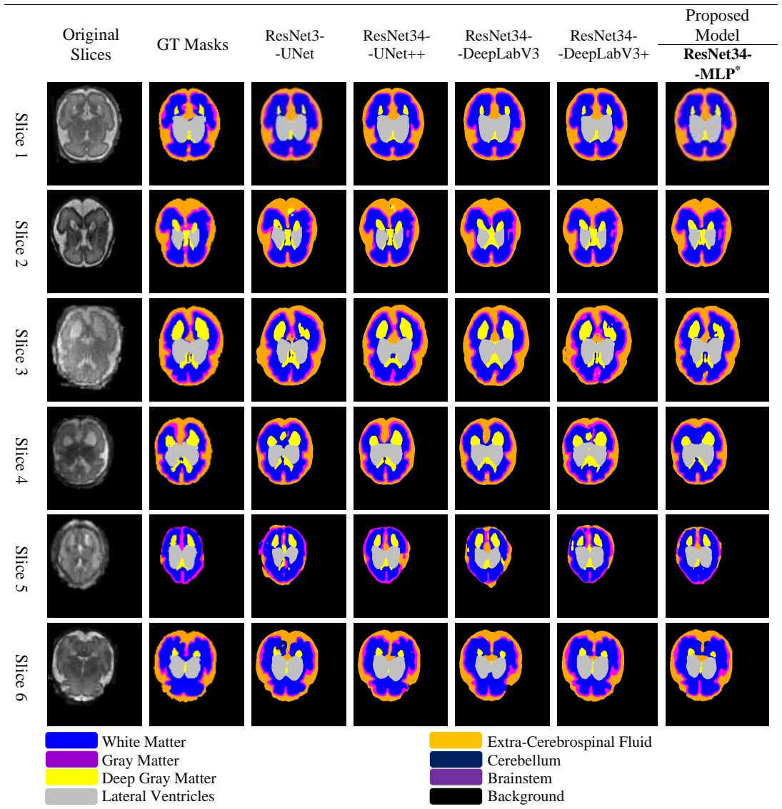
- The architecture outperforms UNet, UNet++, DeepLabV3, and DeepLabV3+ across accuracy, DSC, IoU, and precision on the reported dataset.
- Reduced parameter count and bilinear upsampling produce faster inference times suitable for clinical deployment.
- MLP modules in the decoder improve handling of complex structures such as white matter, ventricles, and cerebellum.
- The design supports integration into real-time prenatal diagnostic workflows.
Where Pith is reading between the lines
- The same encoder-decoder pattern could be tested on other motion-prone medical imaging tasks such as cardiac or abdominal MRI.
- Further reduction of the decoder could enable on-device inference for portable prenatal ultrasound systems.
- Adding uncertainty estimation to the MLP refinement step might help flag low-confidence regions for radiologist review.
Load-bearing premise
The FeTA 2021 dataset and 5-fold cross-validation protocol capture the full range of motion artifacts, intensity inhomogeneities, and gestational-age variability in real clinical fetal MRI scans.
What would settle it
A new test set of fetal MRI scans from different scanners or with higher motion artifacts yielding Dice scores below 90% or IoU below 86%.
Figures
read the original abstract
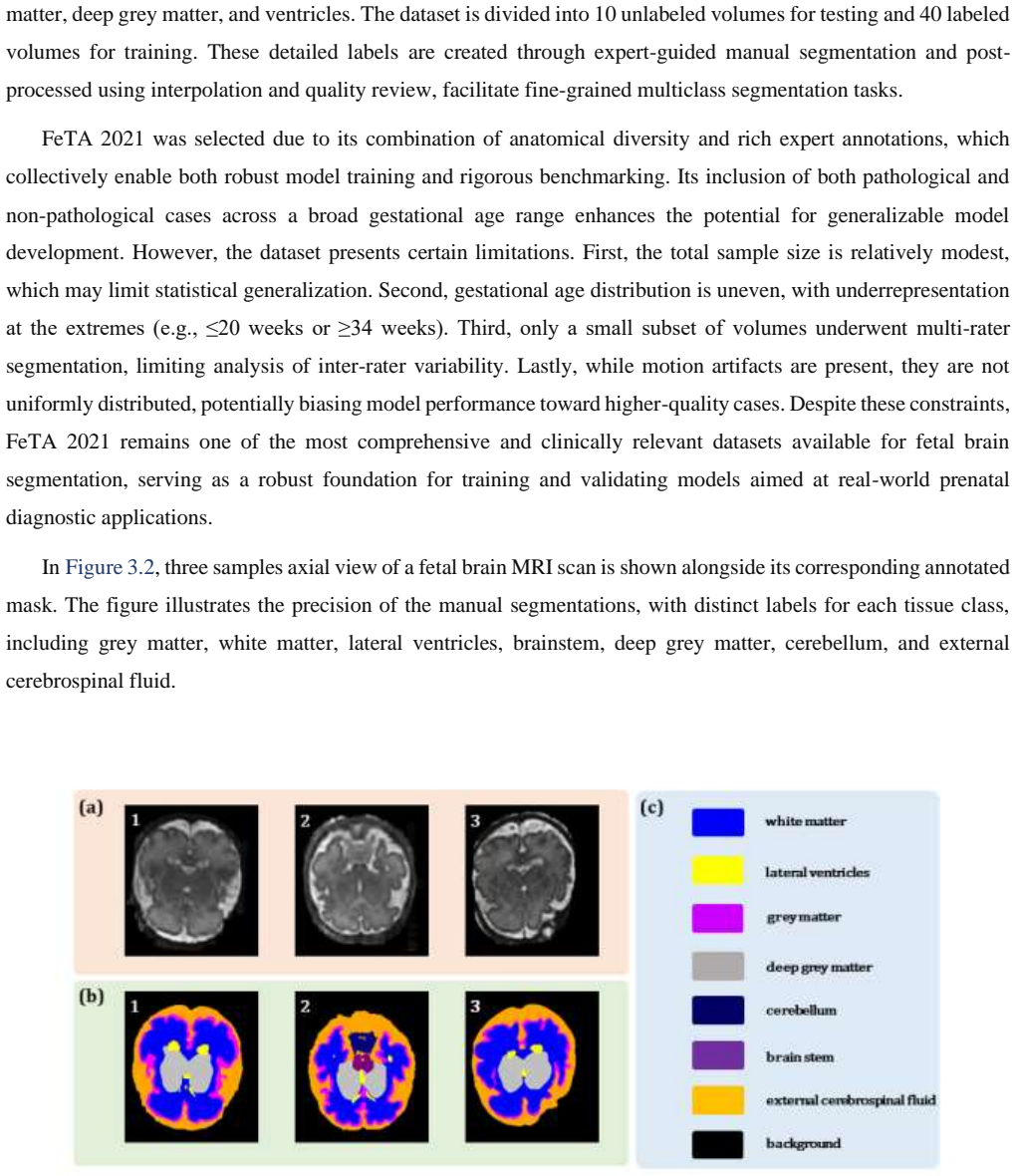
Accurate segmentation of fetal brain tissues in Magnetic Resonance Imaging (MRI) is critical for early diagnosis of congenital abnormalities and improving prenatal care. However, the task remains difficult because of fetal motion, low tissue contrast, and major anatomical variability throughout gestational ages, particularly in segmenting complex structures such as white matter, gray matter, lateral ventricles, deep gray matter, extra-cerebrospinal fluid, cerebellum, and brainstem. As a solution to these difficulties, this research introduces a novel deep learning model that combines a ResNet-34 encoder with a lightweight decoder leveraging multi-layer perceptron (MLP) modules for adaptive feature refinement. This design specifically enhances the model's ability to preserve anatomical boundaries and mitigate segmentation errors caused by motion artifacts and intensity inhomogeneities. Computational efficiency is achieved by reducing parameter count, employing bilinear upsampling instead of transposed convolutions, and optimizing the decoder for speed without sacrificing accuracy. Trained and validated on the FeTA 2021 dataset using 5-fold cross-validation, the proposed model outperforms baseline architectures such as UNet, UNet++, DeepLabV3, and DeepLabV3+, achieving an average Accuracy of 97.37% with a mean Dice Similarity Coefficient (DSC) of 90.33%, mean Intersection over Union (IoU) of 86.93%, and Precision of 90.83%. Additionally, its fast inference time and reduced computational load make it well-suited for integration into real-time clinical workflows.
Editorial analysis
A structured set of objections, weighed in public.
Referee Report
Summary. The manuscript proposes a ResNet-34 encoder combined with a lightweight MLP-based decoder for fetal brain tissue segmentation in MRI. It claims this architecture outperforms UNet, UNet++, DeepLabV3, and DeepLabV3+ on the FeTA 2021 dataset under 5-fold cross-validation, reporting average accuracy of 97.37%, DSC of 90.33%, IoU of 86.93%, and precision of 90.83%, while achieving efficiency via reduced parameters and bilinear upsampling.
Significance. If the performance margins are shown to arise from the proposed architecture under matched training conditions, the work could contribute a computationally lightweight option suitable for clinical fetal MRI workflows. The focus on efficiency and boundary preservation addresses relevant challenges in the domain.
major comments (2)
- [Abstract] Abstract: The headline outperformance claim (DSC 90.33%, IoU 86.93%) is presented without any statement that the baseline models were re-trained from scratch using identical 5-fold splits, data augmentations, optimizer, loss function, and early-stopping criteria as the proposed ResNet-34+MLP model. Without this, the numerical margins cannot be attributed to the architectural modification.
- [Abstract] Abstract: All reported metrics derive from 5-fold cross-validation performed entirely within the FeTA 2021 dataset; no independent held-out test set or external multi-center cohort is mentioned. This protocol does not address generalization across the full range of motion artifacts, intensity inhomogeneities, and gestational-age variability noted in the introduction.
minor comments (1)
- [Abstract] The abstract refers to 'multi-layer perceptron (MLP) modules for adaptive feature refinement' and 'bilinear upsampling instead of transposed convolutions' but supplies no diagram, equation, or parameter count for the decoder, making it impossible to assess the claimed efficiency gains.
Simulated Author's Rebuttal
We thank the referee for the detailed and constructive feedback. We address each major comment below with our responses and indicate where revisions will be made to the manuscript.
read point-by-point responses
-
Referee: [Abstract] Abstract: The headline outperformance claim (DSC 90.33%, IoU 86.93%) is presented without any statement that the baseline models were re-trained from scratch using identical 5-fold splits, data augmentations, optimizer, loss function, and early-stopping criteria as the proposed ResNet-34+MLP model. Without this, the numerical margins cannot be attributed to the architectural modification.
Authors: We appreciate the referee's point on ensuring fair attribution of performance gains. The full manuscript (Sections 3.2 Training Protocol and 4.1 Experimental Setup) specifies that all baseline models (UNet, UNet++, DeepLabV3, DeepLabV3+) were re-trained from scratch using identical 5-fold splits on FeTA 2021, the same data augmentations, Adam optimizer, combined Dice+Cross-Entropy loss, batch size, and early-stopping criteria as the proposed model. We will revise the abstract to explicitly state this protocol so that the reported margins are clearly attributable to the ResNet-34+MLP architecture. revision: yes
-
Referee: [Abstract] Abstract: All reported metrics derive from 5-fold cross-validation performed entirely within the FeTA 2021 dataset; no independent held-out test set or external multi-center cohort is mentioned. This protocol does not address generalization across the full range of motion artifacts, intensity inhomogeneities, and gestational-age variability noted in the introduction.
Authors: We acknowledge that the evaluation relies on 5-fold cross-validation within FeTA 2021 rather than an external held-out cohort. FeTA 2021 is a multi-center dataset spanning 19–39 weeks gestational age with documented variability in motion artifacts, intensity inhomogeneities, and scanner differences; the 5-fold protocol follows the established benchmark standard to permit direct comparison with prior methods. We will add a limitations paragraph in the discussion section explicitly noting the absence of external validation and outlining plans for future multi-center testing. revision: partial
Circularity Check
No significant circularity detected
full rationale
The paper reports an empirical segmentation result obtained by training and evaluating a ResNet-34+MLP model on the FeTA 2021 dataset under 5-fold cross-validation. No derivation chain, mathematical prediction, or first-principles claim is present that reduces to a self-definition, a fitted parameter renamed as a prediction, or a load-bearing self-citation. The performance numbers are direct outputs of the stated training/validation protocol rather than quantities forced by construction from the model architecture itself.
Axiom & Free-Parameter Ledger
Reference graph
Works this paper leans on
-
[1]
Cai, Z., & Zhao, X. -M. (n.d.). Enhancing Generalized Fetal Brain MRI Segmentation using A Cascade Network with Depth - wise Separable Convolution and Attention Mechanism. Cao, H., Wang, Y., Chen, J., Jiang, D., Zhang, X., Tian, Q., & Wang, M. (20 21a). Swin-Unet: Unet-like Pure Transformer for Medical Image Segmentation. Lecture Notes in Computer Science...
-
[2]
https://doi.org/10.1109/OJEMB.2024.3426969 He, K., Zhang, X., Ren, S., & Sun, J. (2015). Deep Res idual Learning for Image Recognition. Proceedings of the IEEE Computer Society Conference on Computer Vision and Pattern Recognition , 2016-December, 770 –778. https://doi.org/10.1109/CVPR.2016.90 Huang, S., Zhang, X., Cui, Z., Zhang, H., Chen, G., & Shen, D ...
-
[3]
https://doi.org/10.3390/S23020655 Huang, X., Liu, Y., Li, Y., Qi, K., Gao, A., Zheng, B., Liang, D., & Long, X. (2023b). Deep Learning -Based Multiclass Brain Tissue Segmentation in Fetal MRIs. Sensors (Basel, Switzerland), 23(2). https://doi.org/10.3390/s23020655 Hutter, J., Slator, P. J., Jackson, L., Gomes, A. D. S., Ho, A., Story, L., O’Muircheartaigh...
work page internal anchor Pith review Pith/arXiv arXiv doi:10.3390/s23020655 2019
-
[4]
https://doi.org/10.1109/QPAIN66474.2025.11171677 Sathyanesan, A., Zhou, J., Scafidi, J., Heck, D. H., Sillitoe, R. V., & Gallo, V. (2019). Emerging connections between cerebellar development, behaviour and complex brain disorders. Nature Reviews Neuroscience 2019 20:5 , 20(5), 298 –313. https://doi.org/10.1038/s41583-019-0152-2 Shen, L., Zheng, J., Lee, E...
-
[5]
S., Chandrasekaran, S., & Krishnakumar, R
https://doi.org/10.3390/s20185097 Singh, V., Sridar, P., Kim, J., Nanan, R., Poornima, N., Priya, S., Reddy, G. S., Chandrasekaran, S., & Krishnakumar, R. (2021). Semantic Segmentation of Cerebellum in 2D Fetal Ultr asound Brain Images Using Convolutional Neural Networks. IEEE Access, 9, 85864–85873. https://doi.org/10.1109/ACCESS.2021.3088946 Ten Donkela...
discussion (0)
Sign in with ORCID, Apple, or X to comment. Anyone can read and Pith papers without signing in.