Sparse-View Lung Nodule Volumetry from Digitally Reconstructed Radiographs via AReT: Anatomy-Regularized TensoRF
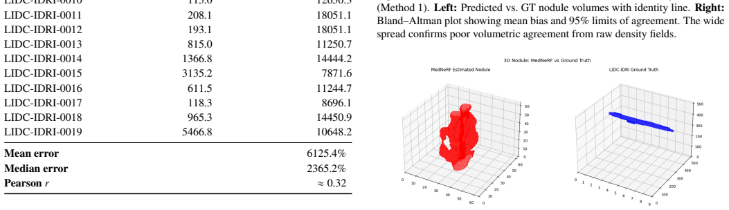
Pith reviewed 2026-06-28 16:46 UTC · model grok-4.3
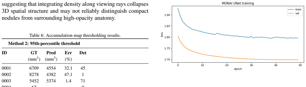
The pith
AReT reconstructs lung nodule volumes from three orthogonal X-ray projections after correcting TensoRF's density shift and adding anatomy-aware regularization.
A machine-rendered reading of the paper's core claim, the machinery that carries it, and where it could break.
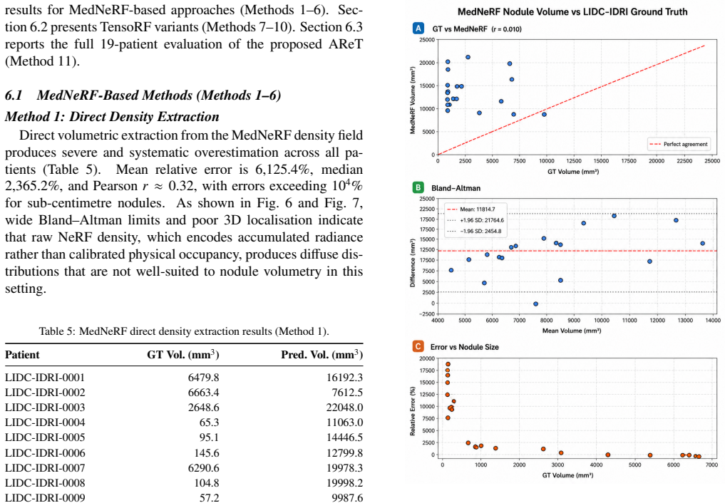
Core claim
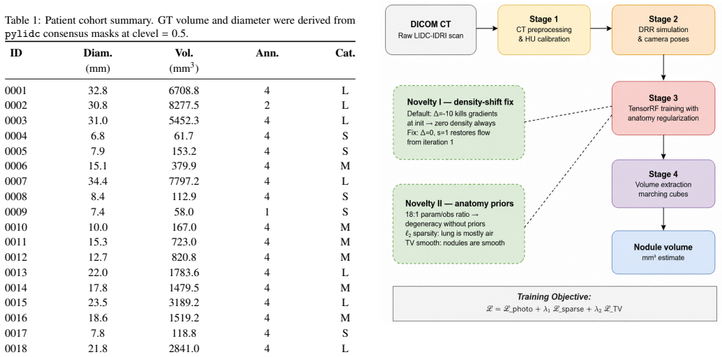
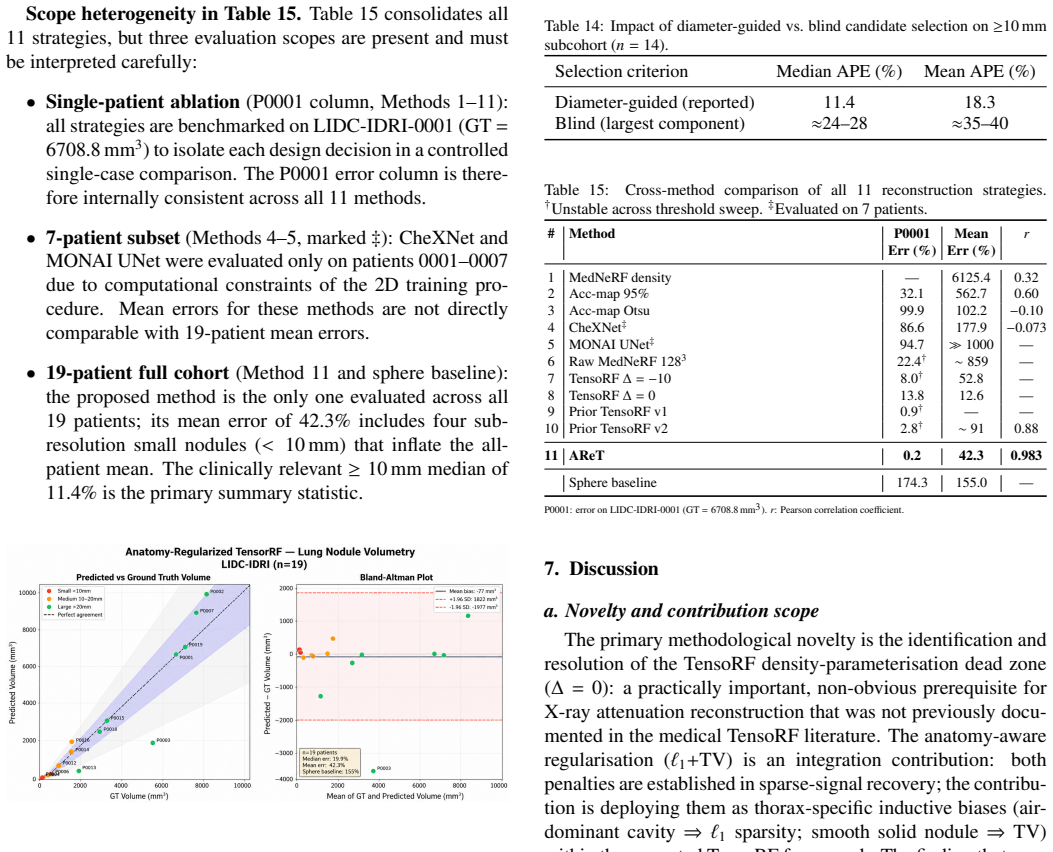
AReT, an anatomy-regularized tensorial radiance field, recovers pulmonary nodule volumes from coronal, sagittal, and axial digitally reconstructed radiographs by first setting the density shift to zero and then enforcing L1 sparsity plus total variation smoothness on the attenuation field, yielding Pearson correlation 0.983, 11.4% median absolute error, and near-zero bias on clinically relevant nodules.
What carries the argument
Anatomy-regularized TensoRF (AReT) that applies L1 sparsity and total variation smoothness to the attenuation field after resetting the density shift from -10 to zero.
If this is right
- Anatomy-aware regularization outperforms generative-prior methods across eleven compared strategies for sparse thoracic imaging.
- Nodule volumes become measurable with median 11.4% error and near-zero bias from three projections alone.
- Spherical volume approximations can be replaced by an 8.4-times more accurate sparse-view method for nodules >=10 mm.
Where Pith is reading between the lines
- The same density-shift correction may unlock other medical attenuation-field tasks that currently fail under default NeRF-style initializations.
- Integration of AReT outputs with existing digital radiography workflows could reduce the number of projections needed for follow-up nodule assessment.
- Extension to four or more projections or to real fluoroscopic sequences would test whether the regularization remains effective under slightly denser but still limited views.
Load-bearing premise
L1 sparsity together with total variation smoothness on the attenuation field is enough to recover accurate nodule volumes from three projections without shape-specific bias across the morphologies in the 19-patient set.
What would settle it
Reconstruction of a new cohort containing a wider range of nodule shapes or attachment patterns that produces median volume error above 20% would falsify the claim.
Figures
read the original abstract
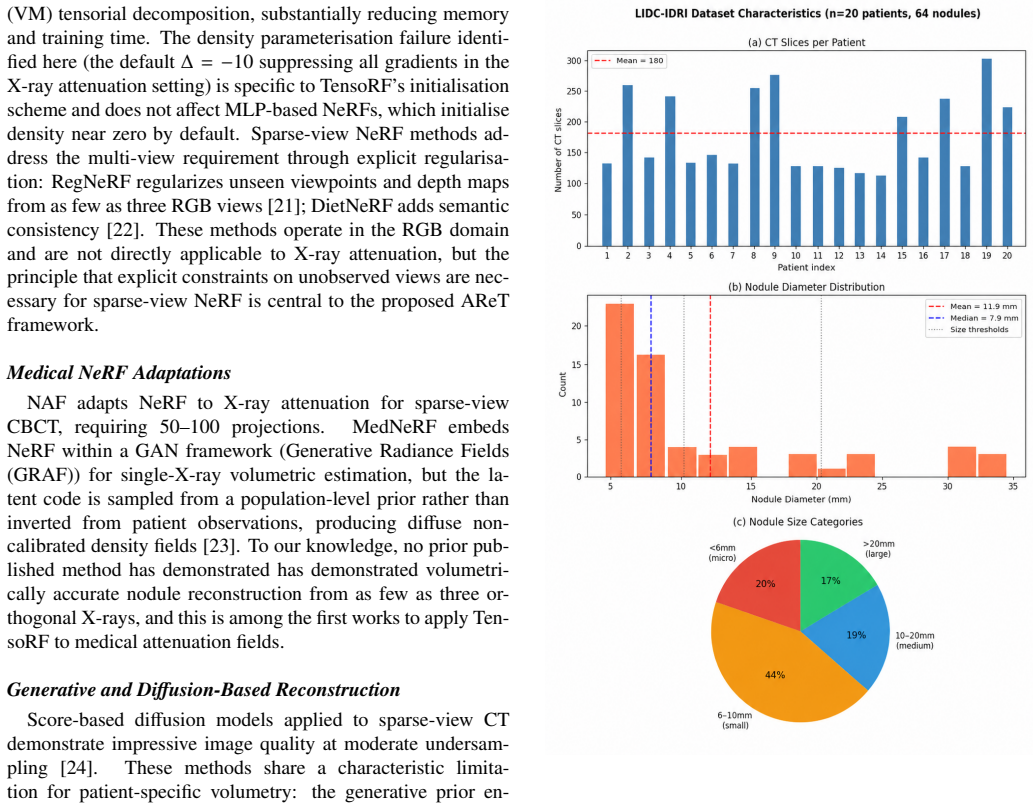
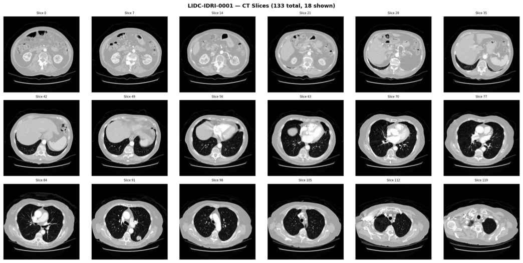
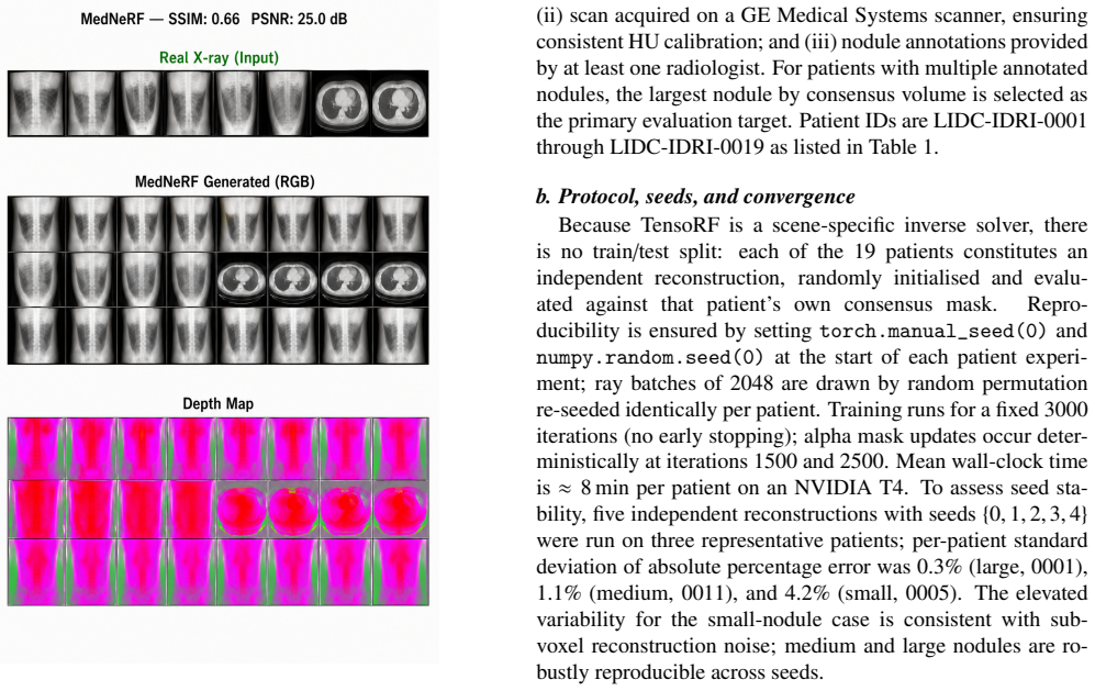
We identify and resolve a previously unreported failure mode in TensoRF when applied to X-ray attenuation fields: the default density shift of -10, originally introduced for RGB scene reconstruction, suppresses density gradients and prevents sparse-view medical reconstruction regardless of learning rate or regularization strategy. Setting the density shift to zero restores gradient flow and enables stable volumetric reconstruction of pulmonary nodules from only three orthogonal X-ray projections. Building on this, we propose AReT, an anatomy-regularized tensorial radiance field framework for lung nodule reconstruction using coronal, sagittal, and axial projections from the LIDC-IDRI dataset (19 patients, radiologist-annotated nodules). Unlike existing NeRF approaches requiring dense multi-view acquisition, AReT is designed for sparse-view thoracic imaging and incorporates chest-anatomy-aware regularization combining L1 sparsity and total variation smoothness. A systematic comparison across 11 reconstruction strategies shows anatomy-aware regularization consistently outperforms generative-prior-guided approaches. Evaluated against radiologist consensus segmentations, AReT achieves Pearson r=0.983 (p<0.0001) for clinically actionable nodules >=10 mm (n=14), median absolute volumetric error of 11.4%, near-zero systematic bias of -77.3 mm^3, and 8.4x improvement over spherical volume approximation.
Editorial analysis
A structured set of objections, weighed in public.
Referee Report
Summary. The paper claims that setting the density shift to zero in TensoRF resolves a failure mode for X-ray attenuation fields, enabling sparse-view reconstruction. They introduce AReT, which adds anatomy-aware L1 and total variation regularization to TensoRF for reconstructing lung nodules from three orthogonal DRRs. On the LIDC-IDRI dataset with 19 patients, AReT achieves Pearson r=0.983 for 14 nodules >=10mm, with 11.4% median absolute error and -77.3 mm^3 bias, outperforming 10 other strategies and spherical approximation by 8.4x.
Significance. If validated, this method could allow accurate nodule volumetry with minimal X-ray projections, reducing patient radiation exposure significantly compared to full CT scans. The use of a public dataset and radiologist annotations, along with systematic comparisons, supports the potential clinical utility of the approach for thoracic imaging.
minor comments (2)
- [Abstract] The total number of nodules in the 19-patient cohort is not stated, only the subset of n=14 for >=10 mm; this should be clarified to assess selection bias.
- [Methods] The specific hyperparameters for the L1 and TV regularization terms (e.g., weighting factors) are not detailed in the provided description, which is important for reproducibility of the anatomy-regularized results.
Simulated Author's Rebuttal
We thank the referee for the positive assessment of our manuscript, the recognition of its potential clinical significance, and the recommendation for minor revision. No specific major comments were provided in the report for us to address point by point.
Circularity Check
No significant circularity; empirical results on external public dataset
full rationale
The paper's central claims consist of an empirical fix (density shift = 0) for TensoRF behavior on attenuation fields plus a new regularization combination (L1 + TV) evaluated via direct comparison to radiologist consensus segmentations on the independent LIDC-IDRI public dataset. No equations reduce a claimed prediction or uniqueness result to a fitted parameter or self-citation defined by the authors; the reported Pearson r, volumetric errors, and bias are measured quantities against external annotations rather than quantities defined by construction from the method itself. The derivation chain therefore remains self-contained against external benchmarks.
Axiom & Free-Parameter Ledger
free parameters (1)
- density shift =
0
axioms (1)
- domain assumption Standard NeRF-style volume rendering integral applies directly to X-ray attenuation coefficients
Reference graph
Works this paper leans on
-
[1]
Global cancer statistics 2020: GLOBOCAN estimates of incidence and mortality worldwide for 36 cancers in 185 countries,
H. Sung, J. Ferlay, R. L. Siegel, M. Laversanne, I. Soerjo- mataram, A. Jemal, and F. Bray, “Global cancer statistics 2020: GLOBOCAN estimates of incidence and mortality worldwide for 36 cancers in 185 countries,”CA Cancer J. Clin., vol. 71, no. 3, pp. 209–249, 2021
2020
-
[2]
Lung-RADS Version 1.1,
American College of Radiology, “Lung-RADS Version 1.1,”ACR Practice Parameter, 2019. Accessed on: Apr. 28, 2026
2019
-
[3]
Guidelines for management of in- cidental pulmonary nodules detected on CT images: from the Fleischner Society 2017,
H. MacMahonet al., “Guidelines for management of in- cidental pulmonary nodules detected on CT images: from the Fleischner Society 2017,”Radiology, vol. 284, no. 1, pp. 228–243, 2017
2017
-
[4]
Is digital chest radiography valid for measurement of pulmonary nodule size?
M. P. Revel, A. Bissery, M. Bienvenu, L. Aycard, C. Lefort, and G. Frija, “Is digital chest radiography valid for measurement of pulmonary nodule size?”Radiology, vol. 232, no. 3, pp. 705–711, 2004
2004
-
[5]
Practical cone-beam algorithm,
L. A. Feldkamp, L. C. Davis, and J. W. Kress, “Practical cone-beam algorithm,”J. Opt. Soc. Am. A, vol. 1, no. 6, pp. 612–619, 1984
1984
-
[6]
Simultaneous algebraic reconstruction technique (SART): a superior implementa- tion of the ART algorithm,
A. H. Andersen and A. C. Kak, “Simultaneous algebraic reconstruction technique (SART): a superior implementa- tion of the ART algorithm,”Ultrason. Imaging, vol. 6, no. 1, pp. 81–94, 1984
1984
-
[7]
NeRF: representing scenes as neural radiance fields for view synthesis,
B. Mildenhall, P. P. Srinivasan, M. Tancik, J. T. Barron, R. Ramamoorthi, and R. Ng, “NeRF: representing scenes as neural radiance fields for view synthesis,” inProc. Eur. Conf. Comput. Vis. (ECCV), Springer, Cham, 2020
2020
-
[8]
NAF: neural attenuation fields for sparse-view CBCT reconstruction,
R. Zha, Y . Zhang, and H. Li, “NAF: neural attenuation fields for sparse-view CBCT reconstruction,” inMed. Im- age Comput. Comput.-Assist. Interv. (MICCAI), Lecture Notes Comput. Sci., Springer, Cham, 2022
2022
-
[10]
TensoRF: tensorial radiance fields,
A. Chen, Z. Xu, A. Geiger, J. Yu, H. Su, and J. Wang, “TensoRF: tensorial radiance fields,” inProc. Eur. Conf. Comput. Vis. (ECCV), Springer, Cham, 2022
2022
-
[11]
The Lung Image Database Con- sortium (LIDC) and Image Database Resource Initiative (IDRI): a completed reference database of lung nodules on CT scans,
S. G. Armatoet al., “The Lung Image Database Con- sortium (LIDC) and Image Database Resource Initiative (IDRI): a completed reference database of lung nodules on CT scans,”Med. Phys., vol. 38, no. 2, pp. 915–931, 2011
2011
-
[12]
A. Corona-Figueroa, J. Frawley, S. Bond-Taylor, S. Bethapudi, H. P. H. Shum, and C. G. Willcocks, “Med- NeRF: Medical Neural Radiance Fields for Reconstruct- ing 3D-aware CT-Projections from a Single X-ray,” in Proc. IEEE Eng. Med. Biol. Soc. (EMBC), pp. 3843–3848, 2022, doi: 10.1109/EMBC48229.2022.9871757
-
[13]
Nonlinear total vari- ation based noise removal algorithms,
L. I. Rudin, S. Osher, and E. Fatemi, “Nonlinear total vari- ation based noise removal algorithms,”Physica D: Non- linear Phenomena, vol. 60, no. 1–4, pp. 259–268, 1992
1992
-
[14]
Image reconstruction in circular cone-beam computed tomography by constrained total- variation minimization,
E. Y . Sidky and X. Pan, “Image reconstruction in circular cone-beam computed tomography by constrained total- variation minimization,”Physics in Medicine&Biology, vol. 53, pp. 4777–4807, 2008
2008
-
[15]
Robust uncertainty principles: exact signal reconstruction from highly incom- plete frequency information,
E. J. Candès, J. Romberg, and T. Tao, “Robust uncertainty principles: exact signal reconstruction from highly incom- plete frequency information,”IEEE Transactions on In- formation Theory, vol. 52, pp. 489–509, 2006
2006
-
[16]
Compressed sensing,
D. L. Donoho, “Compressed sensing,”IEEE Transactions on Information Theory, vol. 52, pp. 1289–1306, 2006
2006
-
[17]
Prior image con- strained compressed sensing (PICCS) for sparse-view CT reconstruction,
G.-H. Chen, J. Tang, and S. Leng, “Prior image con- strained compressed sensing (PICCS) for sparse-view CT reconstruction,”Medical Physics, vol. 35, no. 2, pp. 660– 663, 2008
2008
-
[18]
Maximum likelihood recon- struction for emission tomography,
L. A. Shepp and Y . Vardi, “Maximum likelihood recon- struction for emission tomography,”IEEE Transactions on Medical Imaging, vol. 1, no. 2, pp. 113–122, 1982
1982
-
[19]
Physics- informed neural networks: A deep learning framework for solving forward and inverse problems involving nonlinear partial differential equations,
M. Raissi, P. Perdikaris, and G. E. Karniadakis, “Physics- informed neural networks: A deep learning framework for solving forward and inverse problems involving nonlinear partial differential equations,”Journal of Computational Physics, vol. 378, pp. 686–707, 2019
2019
-
[20]
Learned primal-dual reconstruc- tion,
J. Adler and O. Öktem, “Learned primal-dual reconstruc- tion,”IEEE Transactions on Medical Imaging, vol. 37, no. 6, pp. 1322–1332, 2018
2018
-
[21]
RegNeRF: Regularizing neu- ral radiance fields for view synthesis from sparse inputs,
M. Niemeyer, A. Barron, N. Mildenhall, M. Sajjadi, A. Geiger, and A. Radwan, “RegNeRF: Regularizing neu- ral radiance fields for view synthesis from sparse inputs,” inProc. IEEE/CVF Conf. Comput. Vis. Pattern Recognit. (CVPR), pp. 5480–5490, 2022
2022
-
[22]
Putting NeRF on a diet: Semantically consistent few-shot view synthe- sis,
A. Jain, M. Tancik, and P. Abbeel, “Putting NeRF on a diet: Semantically consistent few-shot view synthe- sis,” inProc. IEEE/CVF Int. Conf. Comput. Vis. (ICCV), pp. 5885–5894, 2021. 18
2021
-
[23]
GRAF: generative radiance fields for 3D-aware image synthesis,
K. Schwarz, A. Sauer, Y . Liao, and A. Geiger, “GRAF: generative radiance fields for 3D-aware image synthesis,” inAdv. Neural Inf. Process. Syst. (NeurIPS), 2020
2020
-
[24]
Score-based diffusion models for accelerated MRI,
H. Chung and J. C. Ye, “Score-based diffusion models for accelerated MRI,”Medical Image Analysis, vol. 80, p. 102479, 2022
2022
-
[25]
Adam: a method for stochas- tic optimization,
D. P. Kingma and J. Ba, “Adam: a method for stochas- tic optimization,” inProc. Int. Conf. Learn. Represent. (ICLR), 2015
2015
-
[26]
A threshold selection method from gray-level histograms,
N. Otsu, “A threshold selection method from gray-level histograms,”IEEE Trans. Syst. Man Cybern., vol. 9, no. 1, pp. 62–66, 1979
1979
-
[27]
Validation, comparison, and combi- nation of algorithms for automatic detection of pulmonary nodules in computed tomography images,
A. A. A. Setioet al., “Validation, comparison, and combi- nation of algorithms for automatic detection of pulmonary nodules in computed tomography images,”Med. Image Anal., vol. 42, pp. 1–13, 2017
2017
-
[28]
Kidney Stone Detection in CT Scans: A Hybrid Ap- proach with Machine Learning and Deep Learning,
V . Kundrapu, T. R. Mettukuru, P. Narisetty, and T. Singh, “Kidney Stone Detection in CT Scans: A Hybrid Ap- proach with Machine Learning and Deep Learning,”IFIP Adv. Inf. Commun. Technol., Springer Nature Switzerland, 2025, doi: 10.1007/978-3-031-98360-3_10
-
[29]
XAI based model evaluation by applying do- main knowledge,
K. S. Srikanth, T. K. Ramesh, S. Palaniswamy, and R. Srinivasan, “XAI based model evaluation by applying do- main knowledge,” inProc. IEEE Int. Conf. Electron. Com- put. Commun. Technol. (CONECCT), IEEE, 2022
2022
-
[30]
Deep learning for enhanced delineation and classification in brain MRI images,
K. Afnaanet al., “Deep learning for enhanced delineation and classification in brain MRI images,”IFIP Adv. Inf. Commun. Technol., Springer Nature Switzerland, 2025, doi: 10.1007/978-3-031-98356-6_11
-
[31]
K. Afnaanet al., “Leveraging convolutional neural net- works for gait recognition and individual identification for improved neurological care,”IFIP Adv. Inf. Com- mun. Technol., Springer Nature Switzerland, 2025, doi: 10.1007/978-3-031-98356-6_10. 19
discussion (0)
Sign in with ORCID, Apple, or X to comment. Anyone can read and Pith papers without signing in.