Justifying Diagnosis Decisions by Deep Neural Networks
Pith reviewed 2026-05-24 22:28 UTC · model grok-4.3
The pith
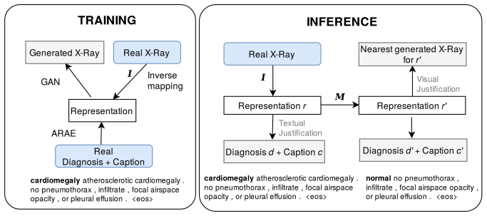
A neural network maps X-ray images to text to produce both a diagnosis and a readable justification.
A machine-rendered reading of the paper's core claim, the machinery that carries it, and where it could break.
Core claim
The authors claim that mapping an X-ray image to a continuous textual representation allows a single network to output a diagnosis together with a human-readable justification and a realistic counterfactual image; on expert ratings from a subset of the Indiana University X-ray collection this justification mechanism significantly outperforms saliency-map methods while matching or exceeding single-task accuracy on diagnosis and captioning.
What carries the argument
Mapping an input X-ray to a continuous textual representation that is decoded into both a diagnosis and its justification text.
If this is right
- Clinicians receive an explicit textual trace they can read to assess whether a diagnosis rests on relevant image features.
- The same network supplies a visual counterfactual showing what an image would look like under the next most likely diagnosis.
- Multi-task training preserves high diagnosis accuracy while producing explanations, removing the need for separate explanation modules.
- The textual output can be inspected or edited by a human before the diagnosis is accepted into the medical record.
Where Pith is reading between the lines
- The same textual-representation step could be applied to other imaging modalities such as CT or MRI to produce comparable justifications.
- If the continuous text space captures diagnostic features reliably, it offers a route to audit or edit model reasoning without retraining the entire vision stack.
- Hospitals could log both the diagnosis and the generated justification text for later review or regulatory reporting.
Load-bearing premise
Expert ratings collected on one hospital data subset will generalize to everyday clinical decisions and the generated text will not add biases that the image alone does not contain.
What would settle it
A follow-up study in which clinicians rate the textual justifications no higher than saliency maps on a broader or more diverse set of cases would falsify the claimed superiority.
Figures


read the original abstract
An integrated approach is proposed across visual and textual data to both determine and justify a medical diagnosis by a neural network. As deep learning techniques improve, interest grows to apply them in medical applications. To enable a transition to workflows in a medical context that are aided by machine learning, the need exists for such algorithms to help justify the obtained outcome so human clinicians can judge their validity. In this work, deep learning methods are used to map a frontal X-Ray image to a continuous textual representation. This textual representation is decoded into a diagnosis and the associated textual justification that will help a clinician evaluate the outcome. Additionally, more explanatory data is provided for the diagnosis by generating a realistic X-Ray that belongs to the nearest alternative diagnosis. With a clinical expert opinion study on a subset of the X-Ray data set from the Indiana University hospital network, we demonstrate that our justification mechanism significantly outperforms existing methods that use saliency maps. While performing multi-task training with multiple loss functions, our method achieves excellent diagnosis accuracy and captioning quality when compared to current state-of-the-art single-task methods.
Editorial analysis
A structured set of objections, weighed in public.
Referee Report
Summary. The manuscript proposes a multi-task deep neural network that encodes frontal chest X-ray images into a continuous textual representation; this representation is decoded to produce both a diagnosis and a textual justification, while an additional decoder generates a realistic X-ray image corresponding to the nearest alternative diagnosis. The central empirical claim is that the textual justification mechanism significantly outperforms saliency-map baselines according to ratings by clinical experts on a subset of the Indiana University hospital X-ray dataset, while the model simultaneously achieves competitive diagnostic accuracy and captioning quality relative to single-task state-of-the-art methods.
Significance. If the expert-study results are reproducible and the textual decoder does not introduce extraneous diagnostic cues, the work would provide a concrete, human-evaluated alternative to saliency-based explanations in medical imaging. The multi-task formulation that reportedly matches or exceeds single-task performance on both diagnosis and captioning is a methodological strength worth noting.
major comments (2)
- [Expert Study section] Expert Study (presumably §4 or §5): the manuscript provides no numerical details on the size of the expert-rated subset, number of raters, blinding protocol, rating scale, inter-rater agreement, or statistical test used to support the claim of “significantly outperforms.” Because this study is the sole evidence offered for the central outperformance claim, the absence of these quantities makes the claim impossible to evaluate.
- [Methods] Methods (textual decoder and multi-task loss): the paper does not report the precise architecture of the continuous textual representation, the weighting schedule for the multiple loss terms, or any ablation that isolates whether the textual decoder adds diagnostic information absent from the raw image. This directly bears on the assumption that the justification is faithful to the input radiograph.
minor comments (2)
- [Abstract] The abstract states “excellent diagnosis accuracy” without reporting the actual accuracy, AUC, or comparison numbers; these should be stated quantitatively even in the abstract.
- [Figures/Tables] Figure captions and table headings should explicitly state the dataset split (train/val/test) and whether the expert study used the same split.
Simulated Author's Rebuttal
We thank the referee for the constructive and detailed review. The comments highlight important omissions that affect the evaluability of the central claims. We address each point below and commit to revisions that will supply the missing information without altering the reported results.
read point-by-point responses
-
Referee: [Expert Study section] Expert Study (presumably §4 or §5): the manuscript provides no numerical details on the size of the expert-rated subset, number of raters, blinding protocol, rating scale, inter-rater agreement, or statistical test used to support the claim of “significantly outperforms.” Because this study is the sole evidence offered for the central outperformance claim, the absence of these quantities makes the claim impossible to evaluate.
Authors: We agree that the Expert Study section is missing the quantitative and procedural details required to evaluate the significance claim. The original manuscript omitted these specifics, which was an error. In the revision we will expand the section to report the exact size of the expert-rated subset, the number of raters, the blinding protocol, the rating scale, inter-rater agreement statistics, and the statistical test used. These additions will make the outperformance result fully evaluable while preserving the study design and outcomes already obtained. revision: yes
-
Referee: [Methods] Methods (textual decoder and multi-task loss): the paper does not report the precise architecture of the continuous textual representation, the weighting schedule for the multiple loss terms, or any ablation that isolates whether the textual decoder adds diagnostic information absent from the raw image. This directly bears on the assumption that the justification is faithful to the input radiograph.
Authors: We concur that the Methods section lacks sufficient specification of the textual decoder architecture, the loss-term weighting schedule, and an ablation isolating the decoder’s contribution to diagnosis. These omissions limit assessment of explanation faithfulness. The revision will include the encoder/decoder layer details and dimensions for the continuous textual representation, the precise weighting schedule used during multi-task training, and a new ablation study that compares diagnostic performance with and without the textual decoder. This will directly address concerns about extraneous diagnostic cues. revision: yes
Circularity Check
No significant circularity; central claim rests on external expert evaluation
full rationale
The paper maps X-ray images to a continuous textual representation via multi-task training, decodes to diagnosis plus justification, and generates alternative X-rays. The key performance claim (outperformance over saliency maps) is supported solely by a clinical expert opinion study on an Indiana University subset, presented as an independent human evaluation. No equations, self-citations, fitted parameters renamed as predictions, or self-definitional steps appear in the abstract or description. The derivation chain is self-contained against external benchmarks and does not reduce to its inputs by construction.
Axiom & Free-Parameter Ledger
free parameters (1)
- multi-task loss weights
Reference graph
Works this paper leans on
- [1]
-
[2]
H.-C. Shin, K. Roberts, L. Lu, D. Demner-Fushman, J. Yao, R. M. Sum- mers, Learning to read chest x-rays: recurrent neural cascade model for automated image annotation, in: Proceedings of the IEEE Conference on Computer Vision and Pattern Recognition, 2016, pp. 2497–2506
work page 2016
-
[3]
M. T. Islam, M. A. Aowal, A. T. Minhaz, K. Ashraf, Abnormality detection and localization in chest x-rays using deep convolutional neural networks, arXiv preprint arXiv:1705.09850 (2017). 19
work page internal anchor Pith review Pith/arXiv arXiv 2017
-
[4]
Interpretation of Mammogram and Chest X-Ray Reports Using Deep Neural Networks - Preliminary Results
H. Salehinejad, J. Barfett, S. Valaee, E. Colak, A. Mnatzakanian, T. Dowdell, Interpretation of mammogram and chest radiograph re- ports using deep neural networks-preliminary results, arXiv preprint arXiv:1708.09254 (2017)
work page internal anchor Pith review Pith/arXiv arXiv 2017
- [5]
-
[6]
I. Goodfellow, J. Pouget-Abadie, M. Mirza, B. Xu, D. Warde-Farley, S. Ozair, A. Courville, Y. Bengio, Generative adversarial nets, Advances in Neural Information Processing Systems (2014) 2672–2680
work page 2014
-
[7]
J.-G. Lee, S. Jun, Y.-W. Cho, H. Lee, G. B. Kim, J. B. Seo, N. Kim, Deep learning in medical imaging: general overview, Korean Journal of Radiology 18 (2017) 570–584
work page 2017
-
[8]
CheXNet: Radiologist-Level Pneumonia Detection on Chest X-Rays with Deep Learning
P. Rajpurkar, J. Irvin, K. Zhu, B. Yang, H. Mehta, T. Duan, D. Ding, A. Bagul, C. Langlotz, K. Shpanskaya, et al., Chexnet: Radiologist-level pneumonia detection on chest x-rays with deep learning, arXiv preprint arXiv:1711.05225 (2017)
work page internal anchor Pith review Pith/arXiv arXiv 2017
-
[9]
L. Oakden-Rayner, Chexnet: an in-depth review, https://lukeoakdenrayner.wordpress.com/2018/01/24/ chexnet-an-in-depth-review/ , 2018
work page 2018
-
[10]
D. Demner-Fushman, M. D. Kohli, M. B. Rosenman, S. E. Shooshan, L. Ro- driguez, S. Antani, G. R. Thoma, C. J. McDonald, Preparing a collection of radiology examinations for distribution and retrieval, Journal of the American Medical Informatics Association 23 (2015) 304–310
work page 2015
-
[11]
L. Oakden-Rayner, Exploring the chestxray14 dataset: prob- lems, https://lukeoakdenrayner.wordpress.com/2017/12/18/ the-chestxray14-dataset-problems/ , 2017
work page 2017
-
[12]
X. Wang, Y. Peng, L. Lu, Z. Lu, M. Bagheri, R. M. Summers, Chestx-ray8: Hospital-scale chest x-ray database and benchmarks on weakly-supervised classification and localization of common thorax diseases, in: Computer Vision and Pattern Recognition (CVPR), 2017 IEEE Conference on, IEEE, 2017, pp. 3462–3471
work page 2017
-
[13]
F. A. Gers, J. Schmidhuber, F. Cummins, Learning to forget: Continual prediction with LSTM, Neural Computation 12 (2000) 2451–2471
work page 2000
-
[14]
B. Jing, P. Xie, E. Xing, On the automatic generation of medical imaging reports, arXiv preprint arXiv:1711.08195 (2017)
work page internal anchor Pith review Pith/arXiv arXiv 2017
- [15]
- [16]
-
[17]
X. Wang, Y. Peng, L. Lu, Z. Lu, R. M. Summers, Tienet: Text-image embedding network for common thorax disease classification and reporting in chest x-rays, in: Proceedings of the IEEE Conference on Computer Vision and Pattern Recognition, 2018, pp. 9049–9058
work page 2018
- [18]
-
[19]
S. Zagoruyko, N. Komodakis, Paying more attention to attention: Improv- ing the performance of convolutional neural networks via attention transfer, arXiv preprint arXiv:1612.03928 (2016)
work page internal anchor Pith review Pith/arXiv arXiv 2016
-
[20]
S. C. Arjovsky, Martin, L. Bottou, Wasserstein gan, arXiv:1701.07875 (2017)
work page internal anchor Pith review Pith/arXiv arXiv 2017
-
[21]
Comparison of Maximum Likelihood and GAN-based training of Real NVPs
I. Danihelka, B. Lakshminarayanan, B. Uria, D. Wierstra, P. Dayan, Com- parison of maximum likelihood and gan-based training of real nvps, arXiv preprint arXiv:1705.05263 (2017)
work page internal anchor Pith review Pith/arXiv arXiv 2017
-
[22]
D. J. Im, H. Ma, G. Taylor, K. Branson, Quantitatively evaluating gans with divergences proposed for training, arXiv preprint arXiv:1803.01045 (2018)
work page internal anchor Pith review Pith/arXiv arXiv 2018
-
[23]
L. M. Radford, Alec, S. Chintala, Unsupervised representation learning with deep convolutional generative adversarial networks, International Conference on Learning Representations (ICLR) (2016)
work page 2016
- [24]
-
[25]
StackGAN: Text to Photo-realistic Image Synthesis with Stacked Generative Adversarial Networks
H. Zhang, T. Xu, H. Li, S. Zhang, X. Huang, X. Wang, D. Metaxas, Stack- gan: Text to photo-realistic image synthesis with stacked generative adver- sarial networks, arXiv preprint arXiv:1612.03242 (2016)
work page internal anchor Pith review Pith/arXiv arXiv 2016
-
[26]
Progressive Growing of GANs for Improved Quality, Stability, and Variation
T. Karras, T. Aila, S. Laine, J. Lehtinen, Progressive growing of gans for improved quality, stability, and variation, arXiv:1710.10196 [cs, stat] (2017). ArXiv: 1710.10196
work page internal anchor Pith review Pith/arXiv arXiv 2017
-
[27]
Y. Choi, M. Choi, M. Kim, J.-W. Ha, S. Kim, J. Choo, Stargan: Unified generative adversarial networks for multi-domain image-to-image trans- lation, IEEE Conference on Computer Vision and Pattern Recognition (CVPR) (2018). 21
work page 2018
-
[28]
J.-Y. Zhu, T. Park, P. Isola, A. A. Efros, Unpaired image-to-image trans- lation using cycle-consistent adversarial networks, in: IEEE International Conference on Computer Vision, 2017
work page 2017
-
[29]
C. Szegedy, W. Zaremba, I. Sutskever, J. Bruna, D. Erhan, I. Goodfel- low, R. Fergus, Intriguing properties of neural networks, International Conference on Learning Representations (ICLR) (2014)
work page 2014
-
[30]
P. Samangouei, M. Kabkab, R. Chellappa, Defense-gan: Protecting clas- sifiers against adversarial attacks using generative models, International Conference on Learning Representations (ICLR) (2018)
work page 2018
-
[31]
T. Che, Y. Li, R. Zhang, R. D. Hjelm, W. Li, Y. Song, Y. Bengio, Maximum-likelihood augmented discrete generative adversarial networks, arXiv preprint arXiv:1702.07983 (2017)
work page internal anchor Pith review Pith/arXiv arXiv 2017
-
[32]
I. Gulrajani, F. Ahmed, M. Arjovsky, V. Dumoulin, A. C. Courville, Im- proved training of wasserstein gans, in: Advances in Neural Information Processing Systems, 2017, pp. 5767–5777
work page 2017
-
[33]
G. Spinks, M.-F. Moens, Generating continuous representations of medical texts, in: Proceedings of the 16th Annual Conference of the North Amer- ican Chapter of the Association for Computational Linguistics: Human Language Technologies, 2018
work page 2018
-
[34]
G. Spinks, M.-F. Moens, Generating text from images in a smooth represen- tation space, in: CLEF2018 Working Notes. CEUR Workshop Proceedings, Avignon, France, 2018, pp. 10–14
work page 2018
-
[35]
G. Spinks, M.-F. Moens, Evaluating textual representations through image generation, in: Proceedings of the 2018 EMNLP Workshop BlackboxNLP: Analyzing and Interpreting Neural Networks for NLP, 2018, pp. 30–39
work page 2018
- [36]
-
[37]
K. Papineni, S. Roukos, T. Ward, W.-J. Zhu, Bleu: a method for au- tomatic evaluation of machine translation, in: Proceedings of the 40th annual meeting on association for computational linguistics, Association for Computational Linguistics, 2002, pp. 311–318
work page 2002
-
[38]
LIN, Rouge: A package for automatic evaluation of summaries, in: Proc
C.-Y. LIN, Rouge: A package for automatic evaluation of summaries, in: Proc. of Workshop on Text Summarization Branches Out, Post Conference Workshop of ACL 2004, 2004
work page 2004
-
[39]
M. Denkowski, A. Lavie, Meteor universal: Language specific translation evaluation for any target language, in: Proceedings of the ninth workshop on statistical machine translation, 2014, pp. 376–380. 22
work page 2014
-
[40]
R. Vedantam, C. Lawrence Zitnick, D. Parikh, Cider: Consensus-based image description evaluation, in: Proceedings of the IEEE conference on computer vision and pattern recognition, 2015, pp. 4566–4575
work page 2015
-
[41]
K. He, X. Zhang, S. Ren, J. Sun, Deep residual learning for image recog- nition, in: Proceedings of the IEEE Conference on Computer Vision and Pattern Recognition, 2016, pp. 770–778. 23 Appendix A. T ext-to-image GAN Architecture Figure A.5 provides an overview of the architecture that was used to train the text-to-image GAN. Figure A.5: Overview of the a...
work page 2016
discussion (0)
Sign in with ORCID, Apple, or X to comment. Anyone can read and Pith papers without signing in.