Echo-POSED: Geometric Self-Distillation for Echocardiography Guidance
Pith reviewed 2026-06-28 17:52 UTC · model grok-4.3
The pith
Echo-POSED learns probe pose from 2D ultrasound slices of 3D volumes by enforcing motion equivariance and cardiac invariance.
A machine-rendered reading of the paper's core claim, the machinery that carries it, and where it could break.
Core claim
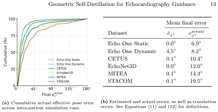
Echo-POSED trains on 2D slices from 3D echocardiography volumes to produce a pose representation that remains geometrically consistent under virtual probe perturbations while ignoring cardiac phase, yielding a combined mean angular error of 8.2 degrees between guided and target views in intra-patient simulations that include cardiac motion on held-out and external vendor-shifted datasets.
What carries the argument
Geometric self-distillation that enforces equivariance to probe motions extracted from 3D volumes while maintaining invariance to cardiac phase, producing an SO(3) x SO(3) pose representation from 2D input images.
If this is right
- The representation maintains geometric consistency when the input view is virtually perturbed.
- It supports both intra-patient and inter-patient guidance simulations that include cardiac motion.
- Performance holds across held-out splits and public external datasets that include vendor shifts.
Where Pith is reading between the lines
- The same slicing-and-distillation pattern could be applied to other 3D-to-2D medical imaging tasks where routine volumetric acquisitions exist.
- Deployment would require checking whether the 8.2-degree error observed in simulation remains acceptable when probe motion is controlled by a human operator in real time.
- The learned representation might also allow offline simulation of entire probe trajectories for sonographer training.
Load-bearing premise
That 2D slices taken from 3D volumes serve as a realistic stand-in for actual probe movements during live scanning and that the learned representation transfers without further labels or tracking.
What would settle it
A test that measures angular error between the method's recommended probe adjustments and simultaneously tracked ground-truth probe poses during actual live 2D echocardiography scans on patients.
Figures


read the original abstract
We introduce Echo-POSED, a self-supervised framework for real-time transthoracic echocardiography (TTE) guidance that recommends probe adjustments directly from 2D ultrasound images, without the need for expert-labelled views or tracked probe trajectories. Instead, it trains on 2D views sliced from routinely acquired 3D echocardiography volumes, enforcing equivariance to probe motions while remaining invariant to cardiac phase, yielding a pose representation on $\mathrm{SO}(3)\times\mathrm{SO}(3)$. Across a held-out split and public external 3D--TTE datasets (including vendor shift), Echo-POSED maintains geometric consistency under virtual perturbations and enables intra- and inter-patient guidance simulations, achieving a combined mean angular error of 8.2 degrees between the guided and target views in intra-patient simulations with cardiac motion.
Editorial analysis
A structured set of objections, weighed in public.
Referee Report
Summary. The paper introduces Echo-POSED, a self-supervised framework for real-time transthoracic echocardiography (TTE) guidance. It trains on 2D views sliced from 3D echocardiography volumes by enforcing equivariance to probe motions (SO(3)×SO(3) pose representation) while remaining invariant to cardiac phase. The method is evaluated on held-out splits and external vendor-shifted 3D-TTE datasets, reporting a combined mean angular error of 8.2° between guided and target views in intra-patient simulations that include cardiac motion.
Significance. If the learned representation transfers to live clinical scanning, the approach could provide a label-free, tracking-free method for probe adjustment in echocardiography, addressing a practical clinical need. The use of routinely acquired 3D volumes for self-supervision and the reported consistency under virtual perturbations are positive aspects, but the absence of direct validation on physical probe trajectories limits the strength of the significance claim.
major comments (2)
- [Abstract] Abstract and evaluation description: the reported 8.2° mean angular error and claims of geometric consistency are obtained exclusively from re-slicing the same 3D volumes used in training (held-out splits and external datasets). No experiments supply tracked real-probe trajectories, acoustic shadowing, or expert-labeled target views on live patients, so the central claim that the representation 'transfers to live clinical scanning' lacks direct supporting evidence.
- [Methods] Methods (loss formulation and data handling): the abstract states a numeric result and claims of equivariance/invariance but supplies no equations for the loss, no definition of the SO(3)×SO(3) representation, no data-exclusion rules, and no error-bar details. This prevents verification of whether the metric supports the stated claims about parameter-free or self-supervised behavior.
minor comments (1)
- [Abstract] Notation: the pose representation is described as lying on SO(3)×SO(3) without an explicit definition of the two rotation groups or how they map to probe orientation versus cardiac phase.
Simulated Author's Rebuttal
We thank the referee for the constructive feedback on our manuscript. We address the major comments point by point below, clarifying the evaluation design and methodological presentation while acknowledging the scope of our current experiments.
read point-by-point responses
-
Referee: [Abstract] Abstract and evaluation description: the reported 8.2° mean angular error and claims of geometric consistency are obtained exclusively from re-slicing the same 3D volumes used in training (held-out splits and external datasets). No experiments supply tracked real-probe trajectories, acoustic shadowing, or expert-labeled target views on live patients, so the central claim that the representation 'transfers to live clinical scanning' lacks direct supporting evidence.
Authors: We agree that all reported results derive from virtual re-slicing of 3D volumes rather than physical probe trajectories acquired on live patients. This design choice enables exact control over SO(3)×SO(3) perturbations and cardiac-phase invariance without additional hardware or annotations, which is central to the self-supervised approach. The 8.2° figure specifically measures intra-patient guidance error under simulated cardiac motion on held-out and external volumes. We will revise the abstract and add a dedicated limitations paragraph to state the evaluation scope more precisely and note that transfer to live scanning remains to be validated in future clinical studies. revision: partial
-
Referee: [Methods] Methods (loss formulation and data handling): the abstract states a numeric result and claims of equivariance/invariance but supplies no equations for the loss, no definition of the SO(3)×SO(3) representation, no data-exclusion rules, and no error-bar details. This prevents verification of whether the metric supports the stated claims about parameter-free or self-supervised behavior.
Authors: The abstract provides a concise overview; the full loss (Eq. 3), SO(3)×SO(3) representation (Sec. 3.1), data-exclusion criteria (Sec. 4.1), and error reporting (mean ± std in Tables 1–3) appear in the Methods and Results sections. The framework is self-supervised because it uses only geometric consistency from slicing and requires no expert labels or tracked trajectories. We will add a short cross-reference in the abstract to the relevant sections for improved readability, but we do not believe equations belong in the abstract itself. revision: no
- Direct experiments with tracked physical probe trajectories, acoustic shadowing, and expert-labeled live-patient views are absent from the study and cannot be supplied without new data collection outside the current 3D-volume dataset.
Circularity Check
No significant circularity: self-supervised training on synthetic slices evaluated on held-out data
full rationale
The paper describes a self-supervised framework that enforces equivariance to probe motions (SO(3)×SO(3)) and invariance to cardiac phase on 2D views sliced from 3D echocardiography volumes. The reported mean angular error of 8.2° is measured on held-out splits and external vendor-shifted datasets under virtual perturbations, which are distinct from the training slices. No component of the pose representation is defined in terms of the evaluation metric by construction, no load-bearing self-citations are invoked, and the derivation does not reduce the guidance simulation results to a fitted input or renaming of the input data. This is a standard self-supervised setup whose central claim remains independently testable on the held-out virtual data.
Axiom & Free-Parameter Ledger
Reference graph
Works this paper leans on
-
[1]
In: Linguraru, M.G., Dou, Q., Feragen, A., Giannarou, S., Glocker, B., Lekadir, K., Schnabel, J.A
Bao, M., Wang, Y., Wei, X., Jia, B., Fan, X., Lu, D., Gu, Y., Cheng, J., Zhang, Y., Wang, C., Zhu, H.: Real-World Visual Navigation for Cardiac Ultrasound View Planning. In: Linguraru, M.G., Dou, Q., Feragen, A., Giannarou, S., Glocker, B., Lekadir, K., Schnabel, J.A. (eds.) Medical Image Computing and Computer As- sisted Intervention – MICCAI 2024. pp. 3...
-
[2]
Bernard, O., Heyde, B., Alessandrini, M., Barbosa, D., Camarasu-Pop, S., Cer- venansky, F., Valette, S., Mirea, O., Galli, E., Geleijnse, M.L., Papachristidis, A., Bosch, J.G., D’hooge, J.: Challenge on Endocardial Three-dimensional Ultra- sound Segmentation (CETUS) (2014),https://lirias.kuleuven.be/retrieve/ 9fc21745-b975-4a45-9027-e3285e805b1b
2014
-
[3]
Bi, Y., Jiang, Z., Duelmer, F., Huang, D., Navab, N.: Machine Learning in Robotic Ultrasound Imaging: Challenges and Perspectives. Annual Review of Control, Robotics, and Autonomous Systems7(1), 335–357 (Jul 2024).https://doi.org/ 10.1146/annurev-control-091523-100042,https://www.annualreviews.org/ content/journals/10.1146/annurev-control-091523-100042
-
[4]
Robots and AI: Illusions and Social Dilemmas
Carnahan, P., Moore, J., Bainbridge, D., Eskandari, M., Chen, E.C.S., Peters, T.M.: DeepMitral: Fully Automatic 3D Echocardiography Segmentation for Pa- tient Specific Mitral Valve Modelling. In: De Bruijne, M., Cattin, P.C., Cotin, S., Padoy, N., Speidel, S., Zheng, Y., Essert, C. (eds.) Medical Image Computing and Computer Assisted Intervention – MICCAI...
-
[5]
Droste, R., Drukker, L., Papageorghiou, A.T., Noble, J.A.: Automatic Probe Movement Guidance for Freehand Obstetric Ultrasound (Jul 2020).https:// doi.org/10.48550/arXiv.2007.04480,http://arxiv.org/abs/2007.04480, arXiv:2007.04480
-
[6]
Frontiers in Cardiovascular Medicine10, 1056055 (Feb 2023)
Ferraz, S., Coimbra, M., Pedrosa, J.: Assisted probe guidance in cardiac ultra- sound: A review. Frontiers in Cardiovascular Medicine10, 1056055 (Feb 2023). https://doi.org/10.3389/fcvm.2023.1056055,https://www.frontiersin. org/articles/10.3389/fcvm.2023.1056055/full
- [7]
-
[8]
Hagendorff, A., Kandels, J., Metze, M., Tayal, B., Stöbe, S.: Valid and Repro- ducible Quantitative Assessment of Cardiac Volumes by Echocardiography in Patients with Valvular Heart Diseases—Possible or Wishful Thinking? Diagnos- tics13(7), 1359 (Apr 2023).https://doi.org/10.3390/diagnostics13071359, https://www.mdpi.com/2075-4418/13/7/1359
-
[9]
He, K., Zhang, X., Ren, S., Sun, J.: Deep Residual Learning for Image Recognition (Dec 2015).https://doi.org/10.48550/arXiv.1512.03385,http://arxiv.org/ abs/1512.03385, arXiv:1512.03385
work page internal anchor Pith review Pith/arXiv arXiv doi:10.48550/arxiv.1512.03385 2015
-
[10]
IEEE trans- actions on bio-medical engineering72(7), 2072–2084 (Jul 2025).https://doi
Huh, J., Klein, P., Funka-Lea, G., Sharma, P., Kapoor, A., Kim, Y.H.: AI-Driven View Guidance System in Intra-Cardiac Echocardiography Imaging. IEEE trans- actions on bio-medical engineering72(7), 2072–2084 (Jul 2025).https://doi. org/10.1109/TBME.2025.3533485
-
[11]
Jiang, H., Li, M., Sun, Z., Jia, N., Sun, Y., Luo, S., Song, S., Huang, G.: Structure- aware World Model for Probe Guidance via Large-scale Self-supervised Pre-train. Geometric Self-Distillation for Echocardiography Guidance 17 In: Gomez, A., Khanal, B., King, A., Namburete, A. (eds.) Simplifying Medical Ultrasound. pp. 58–67. Springer Nature Switzerland,...
-
[12]
Jordan, K., Jin, Y., Boza, V., You, J., Cesista, F., Newhouse, L., Bernstein, J.: Muon: An optimizer for hidden layers in neural networks (2024),https: //kellerjordan.github.io/posts/muon/
2024
-
[13]
Ultrasound in Medicine & Biology49(9), 1996–2005 (Sep 2023).https://doi.org/10.1016/ j
Kim, W.J.C., Beqiri, A., Lewandowski, A.J., Mumith, A., Sarwar, R., King, A., Leeson, P., Lamata, P.: Automated Detection of Apical Foreshorten- ing in Echocardiography Using Statistical Shape Modelling. Ultrasound in Medicine & Biology49(9), 1996–2005 (Sep 2023).https://doi.org/10.1016/ j . ultrasmedbio . 2023 . 05 . 003,https : / / www . sciencedirect ....
1996
-
[14]
Kirkpatrick, J.N., Grimm, R., Johri, A.M., Kimura, B.J., Kort, S., Labovitz, A.J., Lanspa, M., Phillip, S., Raza, S., Thorson, K., Turner, J.: Recommendations for Echocardiography Laboratories Participating in Cardiac Point of Care Cardiac Ultrasound (POCUS) and Critical Care Echocardiography Training: Report from the American Society of Echocardiography....
-
[15]
Lang, R.M., Badano, L.P., Mor-Avi, V., Afilalo, J., Armstrong, A., Ernande, L., Flachskampf, F.A., Foster, E., Goldstein, S.A., Kuznetsova, T., Lancellotti, P., Muraru, D., Picard, M.H., Rietzschel, E.R., Rudski, L., Spencer, K.T., Tsang, W., Voigt, J.U.: Recommendations for Cardiac Chamber Quantification by Echocardio- graphy in Adults: An Update from th...
2015
-
[16]
Lang,R.M.,Badano,L.P.,Tsang,W.,Adams,D.H.,Agricola,E.,Buck,T.,Faletra, F.F., Franke, A., Hung, J., Pérez De Isla, L., Kamp, O., Kasprzak, J.D., Lancel- lotti, P., Marwick, T.H., McCulloch, M.L., Monaghan, M.J., Nihoyannopoulos, P., Pandian, N.G., Pellikka, P.A., Pepi, M., Roberson, D.A., Shernan, S.K., Shirali, G.S., Sugeng, L., Ten Cate, F.J., Vannan, M....
-
[17]
Levinson, J., Esteves, C., Chen, K., Snavely, N., Kanazawa, A., Rostamizadeh, A., Makadia,A.:AnAnalysisofSVDforDeepRotationEstimation(Jun2020).https: //doi.org/10.48550/arXiv.2006.14616,http://arxiv.org/abs/2006.14616, arXiv:2006.14616
-
[18]
net/forum?id=BykcN8siz
Li, Y., Cerrolaza, J.J., Sinclair, M., Hou, B., Alansary, A., Khanal, B., Matthew, J., Kainz, B., Rueckert, D.: Standard Plane Localisation in 3D Fetal Ultrasound Using NetworkwithGeometricandImageLoss.MIDL(Apr2018),https://openreview. net/forum?id=BykcN8siz
-
[19]
In: Frangi, A.F., Schnabel, J.A., Davatzikos, C., Alberola-López, C., Fichtinger, G
Li, Y., Khanal, B., Hou, B., Alansary, A., Cerrolaza, J.J., Sinclair, M., Matthew, J., Gupta, C., Knight, C., Kainz, B., Rueckert, D.: Standard Plane Detection in 3D Fetal Ultrasound Using an Iterative Transformation Network. In: Frangi, A.F., Schnabel, J.A., Davatzikos, C., Alberola-López, C., Fichtinger, G. (eds.) Medical 18 E. Stenhede et al. Image Com...
2018
-
[20]
Springer International Publishing, Cham (2018).https://doi.org/10.1007/ 978-3-030-00928-1_45
2018
-
[21]
Decoupled Weight Decay Regularization
Loshchilov, I., Hutter, F.: Decoupled Weight Decay Regularization (Jan 2019). https://doi.org/10.48550/arXiv.1711.05101,http://arxiv.org/abs/1711. 05101, arXiv:1711.05101
work page internal anchor Pith review Pith/arXiv arXiv doi:10.48550/arxiv.1711.05101 2019
-
[22]
Lyon, A.R., López-Fernández, T., Couch, L.S., Asteggiano, R., Aznar, M.C., Bergler-Klein, J., Boriani, G., Cardinale, D., Cordoba, R., Cosyns, B., Cutter, D.J., De Azambuja, E., De Boer, R.A., Dent, S.F., Farmakis, D., Gevaert, S.A., Gorog, D.A., Herrmann, J., Lenihan, D., Moslehi, J., Moura, B., Salinger, S.S., Stephens, R., Suter, T.M., Szmit, S., Tamar...
-
[23]
Madani, A., Arnaout, R., Mofrad, M., Arnaout, R.: Fast and accurate view classifi- cation of echocardiograms using deep learning. npj Digital Medicine1(1), 6 (Mar 2018).https://doi.org/10.1038/s41746- 017- 0013- 1,https://www.nature. com/articles/s41746-017-0013-1
-
[24]
https://doi.org/10.48550/arXiv.2408.09931,http://arxiv.org/abs/2408
Men, Q., Guo, X., Papageorghiou, A.T., Noble, J.A.: Pose-GuideNet: Automatic Scanning Guidance for Fetal Head Ultrasound from Pose Estimation (Aug 2024). https://doi.org/10.48550/arXiv.2408.09931,http://arxiv.org/abs/2408. 09931, arXiv:2408.09931 Geometric Self-Distillation for Echocardiography Guidance 19
-
[25]
A Dynamic Recursive Unified Internet Design (DRUID),
Men, Q., Zhao, H., Drukker, L., Papageorghiou, A.T., Noble, J.A.: ScanA- head: Simplifying standard plane acquisition of fetal head ultrasound. Medi- cal Image Analysis104, 103614 (Aug 2025).https://doi.org/10.1016/j. media.2025.103614,https://www.sciencedirect.com/science/article/pii/ S1361841525001616
work page doi:10.1016/j 2025
-
[26]
Midtvedt, B., Pineda, J., Skärberg, F., Olsén, E., Bachimanchi, H., Wesén, E., Esbjörner, E.K., Selander, E., Höök, F., Midtvedt, D., Volpe, G.: Single-shot self-supervised object detection in microscopy. Nature Communications13(1), 7492 (Dec 2022).https://doi.org/10.1038/s41467- 022- 35004- y,https: //www.nature.com/articles/s41467-022-35004-y
-
[27]
Mitchell, C., Rahko, P.S., Blauwet, L.A., Canaday, B., Finstuen, J.A., Foster, M.C., Horton, K., Ogunyankin, K.O., Palma, R.A., Velazquez, E.J.: Guidelines for Performing a Comprehensive Transthoracic Echocardiographic Examination in Adults: Recommendations from the American Society of Echocardiography. Jour- nal of the American Society of Echocardiograph...
-
[28]
2011.19.1.1,https://e-jcvi.org/DOIx.php?id=10.4250/jcu.2011.19.1.1
Nakatani, S.: Left Ventricular Rotation and Twist: Why Should We Learn? Journal of Cardiovascular Ultrasound19(1), 1 (2011).https://doi.org/10.4250/jcu. 2011.19.1.1,https://e-jcvi.org/DOIx.php?id=10.4250/jcu.2011.19.1.1
work page doi:10.4250/jcu 2011
-
[29]
Origami-Inspired Soft Twisting Actuator,
Narang, A., Bae, R., Hong, H., Thomas, Y., Surette, S., Cadieu, C., Chaudhry, A., Martin, R.P., McCarthy, P.M., Rubenson, D.S., Goldstein, S., Little, S.H., Lang, R.M., Weissman, N.J., Thomas, J.D.: Utility of a Deep-Learning Algo- rithm to Guide Novices to Acquire Echocardiograms for Limited Diagnostic Use. JAMA Cardiology6(6), 624 (Jun 2021).https://doi...
-
[30]
Ultra- sound in Medicine & Biology49(1), 333–346 (Jan 2023).https://doi.org/10
Pasdeloup, D., Olaisen, S.H., Østvik, A., Sabo, S., Pettersen, H.N., Holte, E., Grenne, B., Stølen, S.B., Smistad, E., Aase, S.A., Dalen, H., Løvstakken, L.: Real- Time Echocardiography Guidance for Optimized Apical Standard Views. Ultra- sound in Medicine & Biology49(1), 333–346 (Jan 2023).https://doi.org/10. 1016/j.ultrasmedbio.2022.09.006
2023
-
[31]
Sabo, S., Pasdeloup, D., Pettersen, H.N., Smistad, E., Østvik, A., Olaisen, S.H., Stølen, S.B., Grenne, B.L., Holte, E., Lovstakken, L., Dalen, H.: Real-time guid- ance by deep learning of experienced operators to improve the standardization of echocardiographic acquisitions. European Heart Journal. Imaging Methods and Practice1(2), qyad040 (Nov 2023).htt...
-
[32]
Sabo, S., Pettersen, H.N., Smistad, E., Pasdeloup, D., Stølen, S.B., Grenne, B.L., Lovstakken, L., Holte, E., Dalen, H.: Real-time guiding by deep learn- ing during echocardiography to reduce left ventricular foreshortening and mea- surement variability. European Heart Journal - Imaging Methods and Practice 1(1), qyad012 (May 2023).https://doi.org/10.1093...
-
[33]
Smistad, E., Ostvik, A., Salte, I.M., Melichova, D., Nguyen, T.M., Haugaa, K., Brunvand, H., Edvardsen, T., Leclerc, S., Bernard, O., Grenne, B., Lovstakken, L.: Real-Time Automatic Ejection Fraction and Foreshortening Detection Using Deep Learning. IEEE transactions on ultrasonics, ferroelectrics, and frequency control 67(12), 2595–2604 (Dec 2020).https:...
-
[34]
Tobon-Gomez, C., De Craene, M., McLeod, K., Tautz, L., Shi, W., Hennemuth, A., Prakosa, A., Wang, H., Carr-White, G., Kapetanakis, S., Lutz, A., Rasche, V., Schaeffter, T., Butakoff, C., Friman, O., Mansi, T., Sermesant, M., Zhuang, X., Ourselin, S., Peitgen, H.O., Pennec, X., Razavi, R., Rueckert, D., Frangi, A.F., Rhode, K.S.: Benchmarking framework for...
-
[35]
Vahanian, A., Beyersdorf, F., Praz, F., Milojevic, M., Baldus, S., Bauersachs, J., Capodanno, D., Conradi, L., De Bonis, M., De Paulis, R., Delgado, V., Freemantle, N., Haugaa, K.H., Jeppsson, A., Jüni, P., Pierard, L., Prendergast, B.P., Sádaba, J.S., Tribouilloy, C., Wojakowski, W.: 2021 ESC/EACTS Guide- lines for the management of valvular heart diseas...
-
[36]
Vukadinovic, M., Ieki, H., Sahashi, Y., Ouyang, D., He, B.: Automated Inter- pretable 2D Video Extraction from 3D Echocardiography (Nov 2025).https: //doi.org/10.48550/arXiv.2511.15946,http://arxiv.org/abs/2511.15946, arXiv:2511.15946
-
[37]
Yeung, P.H., Aliasi, M., Papageorghiou, A.T., Haak, M., Xie, W., Namburete, A.I.L.: Learning to map 2D ultrasound images into 3D space with minimal human annotation. Medical Image Analysis70, 101998 (May 2021).https://doi.org/ 10.1016/j.media.2021.101998,https://www.sciencedirect.com/science/ article/pii/S136184152100044X
-
[38]
https://doi.org/10.48550/arXiv.2504.13065,http://arxiv.org/abs/2504
Yue, Y., Wang, Y., Jiang, H., Liu, P., Song, S., Huang, G.: EchoWorld: Learning Motion-Aware World Models for Echocardiography Probe Guidance (Apr 2025). https://doi.org/10.48550/arXiv.2504.13065,http://arxiv.org/abs/2504. 13065, arXiv:2504.13065
-
[39]
Frontiers in Cardiovascular Medicine9, 1016703 (2022).https://doi
Zhao, D., Ferdian, E., Maso Talou, G.D., Quill, G.M., Gilbert, K., Wang, V.Y., Babarenda Gamage, T.P., Pedrosa, J., D’hooge, J., Sutton, T.M., Lowe, B.S., Legget, M.E., Ruygrok, P.N., Doughty, R.N., Camara, O., Young, A.A., Nash, M.P.: MITEA: A dataset for machine learning segmentation of the left ventricle in 3D echocardiography using subject-specific la...
- [40]
-
[41]
Østvik, A., Smistad, E., Aase, S.A., Haugen, B.O., Lovstakken, L.: Real-Time Standard View Classification in Transthoracic Echocardiography Using Convolu- tional Neural Networks. Ultrasound in Medicine & Biology45(2), 374–384 (Feb 2019).https://doi.org/10.1016/j.ultrasmedbio.2018.07.024
discussion (0)
Sign in with ORCID, Apple, or X to comment. Anyone can read and Pith papers without signing in.